

Anti-Human EGFR Recombinant Antibody (Cetuximab)
CAT#: TAB-003
Recombinant monoclonal antibody to Human EGFR. Cetuximab (Erbitux) is an epidermal growth factor receptor (EGFR) inhibitor used for the treatment of metastatic colorectal cancer and head and neck cancer. Cetuximab is a chimeric (mouse/human) monoclonal antibody given by intravenous infusion.
Published Data
Tested Data
Gene Expression

WB
Figure 1 Phosphoarray analysis of signaling responses to sequential treatments.
(A) HCA7 cells were subjected to different regimens containing 10 μg/ml cetuximab and/or 50 μM oxaliplatin: (1) control medium for 24 hours (white), (2) control medium for 24 hours followed by oxaliplatin for 1 hour (black), (3) cetuximab for 24 hours followed by oxaliplatin for 1 hour (red), or (4) oxaliplatin for 24 hours followed by cetuximab for 1 hour (green). Phosphorylation profiles were analyzed using Proteome Profiler Human Phospho-Kinase Array Kit and quantified by densitometry. Mean +/− range is shown for four selected proteins. All results are shown in Suppl. Fig. 3. (B) HCA7 cells were subjected to different regimens containing or not 10 μg/ml cetuximab and/or 50 μM oxaliplatin as indicated in the figure. The expression or phosphorylation of selected proteins was analyzed by Western blotting. Full-length blots are presented in Supplementary Fig. 8. (C) Apoptosis of HCA7 cells was analyzed by annexin V staining after treating the cells first with or without 200 μM of the STAT3 inhibitor S31-201 for 8 hours followed by 18 hour treatment with or without 10 μg/ml cetuximab.
Narvi, E., Vaparanta, K., Karrila, A., Chakroborty, D., Knuutila, S., Pulliainen, A.,... & Elenius, K. (2018). Different responses of colorectal cancer cells to alternative sequences of cetuximab and oxaliplatin. Scientific reports, 8(1), 16579.

Block
Figure 2 Block result.
(A) Schematic representation of hypothetical cellular responses to the opposite sequences of cetuximab and oxaliplatin. Left side: When cetuximab is administered first, it promotes arrest in the G1 phase of the cell cycle and stimulates apoptosis by blocking EGFR-activated survival pathways. The subsequent administration of oxaliplatin (ox) is largely ineffective due to the G1-arrest. Right side: When oxaliplatin is administered first, it actively induces DNA damage provoking apoptosis, as well as DNA repair that partially compensates for the damage. In the absence of cetuximab, the cell cycle is not arrested at G1 allowing for full activity of oxaliplatin. However, oxaliplatin-induced DNA damage itself does eventually cause cell cycle arrest by activating checkpoints in the S and G2 phases (not indicated in the figure). When cetuximab is then added after oxaliplatin, apoptosis is enhanced compared to all other regimens, as the apoptotic effect of both drugs are active, and also, as cetuximab may be suppressing the DNA repair mechanisms caused by oxaliplatin-induced DNA damage. (B) HCA7, DLD-1 and RKO cells were arrested in G1 by 8 hour treatment with or without 3 µM of the CDK4/6 inhibitor abemaciclib followed by 18 hour treatment with or without 50 μM oxaliplatin.(C) Flow cytometry analysis of cell surface EGFR after 24 hour treatment with or without 10 μg/ml cetuximab or 50 μM oxaliplatin.
Narvi, E., Vaparanta, K., Karrila, A., Chakroborty, D., Knuutila, S., Pulliainen, A.,... & Elenius, K. (2018). Different responses of colorectal cancer cells to alternative sequences of cetuximab and oxaliplatin. Scientific reports, 8(1), 16579.

Activ
Figure 3 Sequence of administration determines the effect of the regimen including cetuximab and oxaliplatin.
(A) Representative MTT experiments of HCA7 (left panel) and DLD-1 (right panel) cells. Mean +/− SD is shown. (B) Oxaliplatin ED50 values of sequential treatments from 3–4 independent experiments with HCA7 and DLD-1 cells. (C) Oxaliplatin ED50 values of sequential treatments from single independent experiments with the seven indicated cell lines. (D) HCA7 (left panel) and DLD-1 (right panel) cells suspended in soft agar were (1) treated for 48 hours (for days 4 to 5 of each treatment cycle) with oxaliplatin alone (black curves), or (2) in simultaneous combination with cetuximab (grey curves), (3) treated first for 72 hours (days 1 to 3) with cetuximab followed by 48 hour (days 4 to 5) treatment with oxaliplatin (red curves), or (4) treated first for 48 hours (days 4 to 5) with oxaliplatin followed by 72 hour (days 6 to 8) treatment with cetuximab (green curves). The cycle was repeated three times every 21 days as indicated by black dots.(E) Nude mice carrying HT-29 cell xenografts were treated with i.p. injections of (1) vehicle alone on day 1 of each treatment cycle (white dots), (2) oxaliplatin alone on day 1 (black dots), (3) cetuximab on day 1 followed by oxaliplatin on day 2 (red dots), or (4) oxaliplatin on day 1 followed by cetuximab on day 2 (green dots). The cycle was repeated three times every seven days as indicated by black dots. The concentrations used for oxaliplatin and cetuximab were 10 mg/kg and 40 mg/kg, respectively.
Narvi, E., Vaparanta, K., Karrila, A., Chakroborty, D., Knuutila, S., Pulliainen, A.,... & Elenius, K. (2018). Different responses of colorectal cancer cells to alternative sequences of cetuximab and oxaliplatin. Scientific reports, 8(1), 16579.

Activ
Figure 4 Sequential administration of cetuximab after oxaliplatin reduces G1 arrest and enhances apoptosis.
Cell cycle (A) and apoptosis (B) analyses of HCA7 cells subjected to different regimens containing 10 μg/ml cetuximab and/or 50 μM oxaliplatin: (1) control medium for 24 hours (white), (2) cetuximab for 24 hours (blue), (3) oxaliplatin for 24 hours (black), (4) simultaneous combination of oxaliplatin and cetuximab for 24 hours (grey), or treated sequentially (5) first with cetuximab for 24 hours followed by oxaliplatin for 24 hours (red) or (6) first with oxaliplatin for 24 hours followed by cetuximab for 24 hours (green). Cell cycle was analyzed by PI staining (A) and apoptosis by annexin V staining (B). Mean +/− SD is shown.
Narvi, E., Vaparanta, K., Karrila, A., Chakroborty, D., Knuutila, S., Pulliainen, A.,... & Elenius, K. (2018). Different responses of colorectal cancer cells to alternative sequences of cetuximab and oxaliplatin. Scientific reports, 8(1), 16579.

Inhib
Figure 5 Low concentrations of HT and cetuximab reduce cell growth in colorectal cancer.
HT-29 (A), and WiDr (D) cells were exposed to increasing HT concentrations in presence/absence of EGF (5 ng/ml) for 48 h. Cell viability values, reported as absorbance at 540 nm, were obtained by MTT assay. HT-29 (B), and WiDr (E) cells were exposed to increasing cetuximab concentrations in presence/absence of EGF (5 ng/ml). Values obtained as in A. Numbers represent mean ± DS of three experiments run in triplicate. HT-29 (C), and WiDr (F) cells were exposed to HT (10 μM) and/or cetuximab (1 μg/ml) in presence/absence of EGF (5 ng/ml) for 48 h. These concentrations were used throughout this work, unless otherwise noted. Values obtained as in A.
Terzuoli, E., Nannelli, G., Frosini, M., Giachetti, A., Ziche, M., & Donnini, S. (2017). Inhibition of cell cycle progression by the hydroxytyrosol–cetuximab combination yields enhanced chemotherapeutic efficacy in colon cancer cells. Oncotarget, 8(47), 83207.

FuncS
Figure 6 Combination of low concentrations of HT and cetuximab reduces colony formation of colorectal cancer cells.
Colony formation capability of HT-29 (A), and WiDr (B) cells in response to HT (10 μM) and/or cetuximab (1μg/ml) in presence/absence of EGF (5 ng/ml). Colonies (>75 cells) with 50% efficiency were counted. Results are expressed as surviving factor (SF, see material and methods). ** P <0.01, *** P <0.001, vs. untreated cells. # P <0.05, ### P <0.001 vs. EGF-treated cells.
Terzuoli, E., Nannelli, G., Frosini, M., Giachetti, A., Ziche, M., & Donnini, S. (2017). Inhibition of cell cycle progression by the hydroxytyrosol–cetuximab combination yields enhanced chemotherapeutic efficacy in colon cancer cells. Oncotarget, 8(47), 83207.

IF
Figure 7 EGFR expression in colorectal cancer cells treated with HT and cetuximab alone or combined.
HT-29 (A) and WiDr (B) cells were exposed to low concentration of HT or cetuximab alone or in combination for 8 h and EGFR proteins was analyzed by western blot. Images of immunostaining in HT-29 (C) and WiDr (D) for EGFR (green) and DAPI (blue) in tumor cells exposed to 10 % FBS (a), cetuximab 1 μg/ml (b), HT 10 μM (c), cetuximab 1 μg/ml + HT 10 μM (d), cetuximab 10 μg/ml (e), HT 100 μM (f). Confocal images were captured with Leica SP5 confocal using 63x objective, scale bars 20 μm. HT-29 (E) and WiDr (F) cells were exposed to high concentration of HT or cetuximab for 8 h and EGFR proteins were analyzed by western blot. β-actin has been used to normalized loading (in Figures 3 and 7).
Terzuoli, E., Nannelli, G., Frosini, M., Giachetti, A., Ziche, M., & Donnini, S. (2017). Inhibition of cell cycle progression by the hydroxytyrosol–cetuximab combination yields enhanced chemotherapeutic efficacy in colon cancer cells. Oncotarget, 8(47), 83207.

FC
Figure 8 Cell cycle analysis in cancer cells treated with low concentration of HT and cetuximab combined.
HT-29 (A), and WiDr (B) cells were exposed to HT or cetuximab alone or in combination in presence or absence of EGF for 48 h. The percentage of cells at each stage of the cell cycle was analyzed by flow cytometry after DNA staining with propidium iodide. Quantification of cells residing in G0 and G1 for HT-29 (C), and WiDr (D) are reported. Percent of HT-29- (C), and WiDr-cells (D) in sub Go/G1 phase.
Terzuoli, E., Nannelli, G., Frosini, M., Giachetti, A., Ziche, M., & Donnini, S. (2017). Inhibition of cell cycle progression by the hydroxytyrosol–cetuximab combination yields enhanced chemotherapeutic efficacy in colon cancer cells. Oncotarget, 8(47), 83207.

WB
Figure 9 HT and cetuximab combination modulate the cell cycle checkpoint proteins in colorectal cancer cells.
HT-29 (A, B, C), and WiDr (D, E, F) cells were exposed to HT or cetuximab, alone or in combination in presence of EGF for 48 h and the cell cycle checkpoint proteins were analyzed by western blot.
Terzuoli, E., Nannelli, G., Frosini, M., Giachetti, A., Ziche, M., & Donnini, S. (2017). Inhibition of cell cycle progression by the hydroxytyrosol–cetuximab combination yields enhanced chemotherapeutic efficacy in colon cancer cells. Oncotarget, 8(47), 83207.

IF
Figure 10 HT and cetuximab combination induces caspace3-independent apoptosis in colorectal cancer cells.
Phosphatidylserine (red) and DAPI (blu) exposure, assessed by immunofluorescence, in HT-29 (A, C for quantification) and WiDr (B, D for quantification) cells treated for 48 h with 10% FBS (Ctr) (a), cetuximab 1 μg/ml (b), HT HT 10 μM (c) or cetuximab 1 μg/ml+ HT 10 μM. Confocal images were captured with Leica SP5 confocal using 40 x objective, scale bars 60 μm.*P<0.05; **P <0.01 vs. untreated cells; §P<0.05; §§P<0.01 vs. HT- or cetuximab-treated cells. HT-29 (E), and WiDr (F) cells were exposed to HT or cetuximab alone or in combination for 8 h and caspase-3 activity were analyzed by western blot.
Terzuoli, E., Nannelli, G., Frosini, M., Giachetti, A., Ziche, M., & Donnini, S. (2017). Inhibition of cell cycle progression by the hydroxytyrosol–cetuximab combination yields enhanced chemotherapeutic efficacy in colon cancer cells. Oncotarget, 8(47), 83207.

FuncS
Figure 11 HT-cetuximab combination reduces side effects of cetuximab treatment in colorectal cancer.
CCD-18Co (A), differentiated CaCo2 (B) and HaCaT (C) cells were exposed to the indicated concentration of HT and cetuximab in presence/absence of EGF (5 ng/ml) for 48 h. Cell survival values, reported as absorbance at 540 nm, were obtained by MTT assay. *P<0.05; **P<0.01 vs. untreated cells; §P<0.05; §§P<0.01 vs. cetuximab 10 μg/ml alone; çP<0.05, ççP<0.01 vs EGF-treated cells; #P<0.05, ##P<0.01 vs cetuximab 10 μg/ml plus EGF-treated cells. CCD-18Co (D, upper panel) and differentiated CaCo2 (D, bottom panel) cells were exposed to HT or cetuximab alone or in combination for 8 h and EGFR proteins was analyzed by western blot. CCD-18Co (E) and differentiated CaCo2 (F) cells were exposed to cetuximab (10 μg/ml) alone for 8 h and EGFR proteins were analyzed by western blot. β-actin has been used to normalize loading.
Terzuoli, E., Nannelli, G., Frosini, M., Giachetti, A., Ziche, M., & Donnini, S. (2017). Inhibition of cell cycle progression by the hydroxytyrosol–cetuximab combination yields enhanced chemotherapeutic efficacy in colon cancer cells. Oncotarget, 8(47), 83207.
❮


 ❯
❯

ELISA
Figure 1 Anti-Human EGFR Antibody (TAB-003) in ELISA
ELISA analysis of TAB-003 was performed with EGFR Protein, Human, Recombinant (Isoform vIII, His Tag).
The secondary antibody: HRP-Anti-Human IgG (H+L)

WB
Figure 2 Anti-Human EGFR Antibody (TAB-003) in WB
Western blot analysis of TAB-003 was performed with EGFR Protein, Human, Recombinant (Isoform vIII, His Tag).
TAB-003 incubation concentration: 2ng/μL.
The secondary antibody: HRP-Anti-Human IgG (H+L)
Lane 1: Reducing antigen (0.6μg)

DB
Figure 3 Anti-Human EGFR Antibody (TAB-003) in DB
Dot blot analysis of TAB-003 was performed with EGFR Protein, Human, Recombinant (Isoform vIII, His Tag).
TAB-003 incubation concentration: 2ng/μL.
NC: Negative control PC: Positive control
❮


 ❯
❯

Subcellular Location and Protein Expression
Figure 1 IF staining of human cell line A-431
Immunofluorescent staining of human cell line A-431 shows localization to plasma membrane & cell junctions.
* Image credit: Image credit: Human Protein Atlas https://v21.proteinatlas.org/images/18530/144_E9_2_selected.jpg

Subcellular Location and Protein Expression
Figure 2 IF staining of human cell line U-251 MG
Immunofluorescent staining of human cell line U-251 MG shows localization to plasma membrane.
* Image credit: Image credit: Human Protein Atlas https://v21.proteinatlas.org/images/18530/143_E9_1_blue_red_green.jpg

Normal Tissue
Figure 3 Colon
Glandular cells
Staining:Medium
Intensity: Moderate
Quantity:>75%
Location: Cytoplasmic/membranous
* Image credit: Image credit: Human Protein Atlas https://v21.proteinatlas.org/images/18530/41191_A_9_3.jpg

Normal Tissue
Figure 4 Liver
Cholangiocytes
Staining:Medium
Intensity: Moderate
Quantity: 75%-25%
Location: Cytoplasmic/membranous
* Image credit: Image credit: Human Protein Atlas https://v21.proteinatlas.org/images/18530/41191_A_7_4.jpg

Normal Tissue
Figure 5 Kidney
Bowman's capsule
Staining:Medium
Intensity: Moderate
Quantity: 75%-25%
Collecting ducts
Staining:Medium
Intensity: Strong
Quantity: <25%
Distal tubules
Staining:Medium
Intensity: Strong
Quantity: <25%
Proximal tubules (cell body)
Staining:Medium
Intensity: Strong
Quantity: <25%
* Image credit: Image credit: Human Protein Atlas https://v21.proteinatlas.org/images/18530/41191_A_9_5.jpg

Normal Tissue
Figure 6 Testis
Leydig cells
Staining:Medium
Intensity: Moderate
Quantity:>75%
Pachytene spermatocytes
Staining:Medium
Intensity: Moderate
Quantity: 75%-25%
Round or early spermatids
Staining:Medium
Intensity: Moderate
Quantity: 75%-25%
* Image credit: Image credit: Human Protein Atlas https://v21.proteinatlas.org/images/18530/41191_A_6_6.jpg

Normal Tissue
Figure 7 Placenta
Cytotrophoblasts
Staining:High
Intensity: Strong
Quantity:>75%
Decidual cells
Staining:Medium
Intensity: Moderate
Quantity:>75%
Hofbauer cells
Staining:High
Intensity: Strong
Quantity:>75%
Syncytiotrophoblasts - cell body
Staining:High
Intensity: Strong
Quantity:>75%
Syncytiotrophoblasts - microvilli
Staining:High
Intensity: Strong
* Image credit: Image credit: Human Protein Atlas https://v21.proteinatlas.org/images/18530/41191_A_1_7.jpg

Normal Tissue
Figure 8 Lymph node
Non-germinal center cells
Staining:Medium
Intensity: Strong
Quantity: <25%
Location: Cytoplasmic/membranous
* Image credit: Image credit: Human Protein Atlas https://v21.proteinatlas.org/images/18530/41191_A_7_8.jpg

RNA Expression
Figure 9 RNA cell line category: Cell line enriched (A-431)
Cell lines ordered by descending RNA expression order.
* Image credit: Image credit: Human Protein Atlas https://v21.proteinatlas.org/ENSG00000146648-EGFR
❮


 ❯
❯
Specifications
- Immunogen
- CHO cells transfected with a plasmid bearing a truncated form of EGFR cDNA
- Host Species
- Human
- Derivation
- Chimeric (mouse/human)
- Type
- IgG1 - kappa
- Specificity
- Tested positive against native human antigen
- Species Reactivity
- Human
- Applications
- IF, IP, Neut, FuncS, ELISA, FC, ICC, WB, Block, Activ, Inhib
- Trade name
- erbitux
- CAS
- 205923-56-4
- Generic Name
- Cetuximab
- Biological Half-Life
- 114 hrs
- ATC Code
- L01XC06
- DrugBank
- DB00002
- UNII
- PQX0D8J21J
- ChEMBL
- CHEMBL1201577
- MW
- 145,781.6 g/mol
- Related Disease
- Colorectal cancers, metastatic (EGFR positive)
Product Property
- Purity
- >95.0% as determined by analysis by SDS-PAGE.
- Storage
- Store the antibody (in aliquots) at -20°C. Avoid repeated freezing and thawing of samples.
Applications
- Application Notes
- The EGFR antibody has been reported in applications of IF, IP, Neut, FuncS, ELISA, FC, ICC, WB, Block, Activ, Inhib.
Target
REVIEWS AND Q&AS
CITATIONS
RESOURCES
DOWNLOADS
RELATED PRODUCTS
Inquiry
Navs
Customer Review

There are currently no Customer reviews or questions for TAB-003. Click the button above to contact us or submit your feedback about this product.

Excellent Functional EGFR Ab
We use this anti-EGFR antibody (TAB-003) for functional studies (FuncS) in our cancer cell models. As a chimeric (Human-Mouse) IgG1, it performs reliably. Its broad validation for other methods like WB, FC, and IF makes it an extremely versatile and valuable reagent.
Reliable for IHC and IF
This is our go-to antibody for imaging human EGFR. It works consistently for Immunofluorescence (IF). It's a CHO-expressed chimeric IgG1, and we appreciate that we can use the same antibody for these applications as well as for Western Blot (WB).
Q&As
-
Is TAB-003 suitable for functional studies and other common assays?
A: Yes, TAB-003 is validated for functional studies (FuncS), making it ideal for cancer cell models. It is a chimeric (Human-Mouse) IgG1 and also has broad validation for other key applications, including Western Blot (WB), Flow Cytometry (FC), and ELISA, making it a very versatile reagent.
-
Can I use this antibody for imaging human EGFR, and what is its composition?
A: Absolutely. This antibody is a reliable choice for imaging, as it's confirmed to work consistently for Immunofluorescence (IF) on cells. It is a chimeric (Human-Mouse) IgG1 antibody that is recombinantly produced in a CHO expression system, ensuring high consistency.
View the frequently asked questions answered by Creative Biolabs Support.
Cite This Product
To accurately reference this product in your publication, please use the following citation information:
(Creative Biolabs Cat# TAB-003, RRID: AB_3111731)
Copy citation
Submit Your Publication
Published with our product? Submit your paper and receive a 10% discount on your next order! Share your research to earn exclusive rewards.
Biosimilar Overview
Please refer to Cetuximab Overview to learn more about the mechanism of action, clinical projects, and approved drugs of Cetuximab.
Related Diseases
 Bladder Cancer
 Non-small Cell Lung Cancer
 Pancreatic Cancer
 Hepatocellular Carcinoma
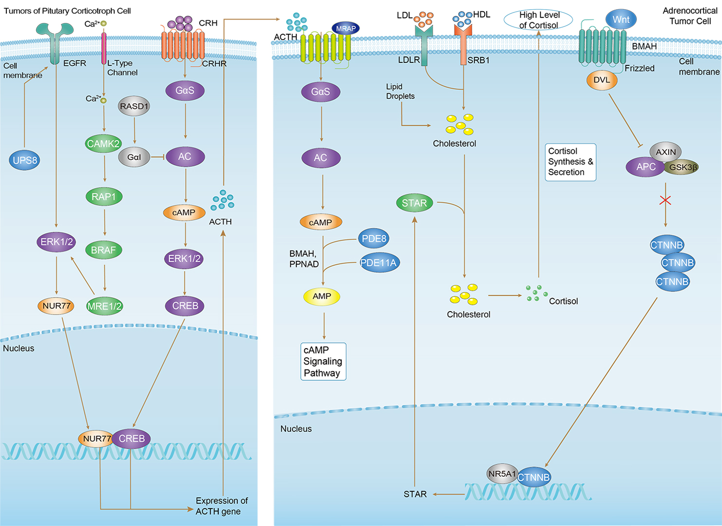 Cushing Syndrome
Related Signaling Pathways
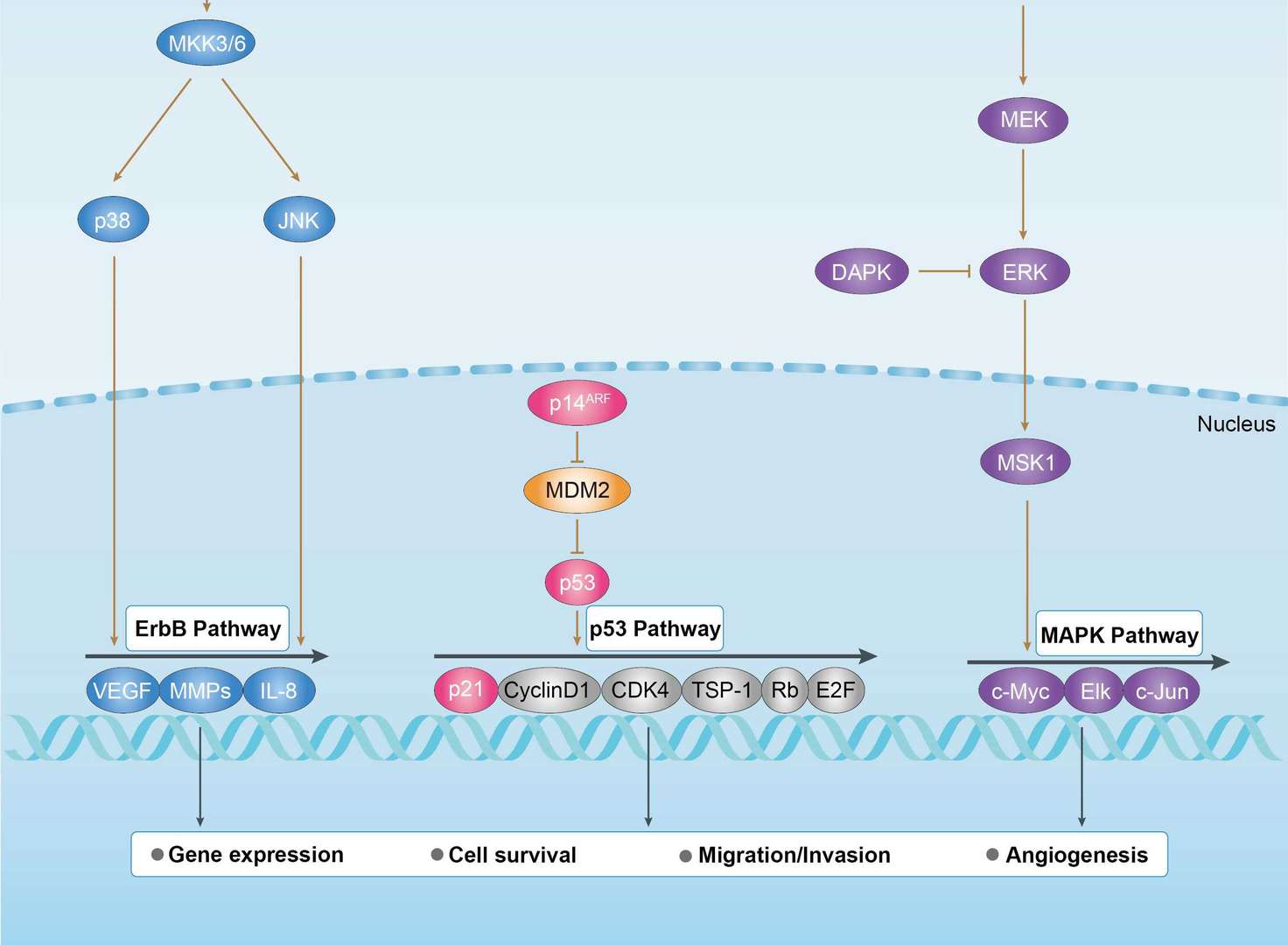
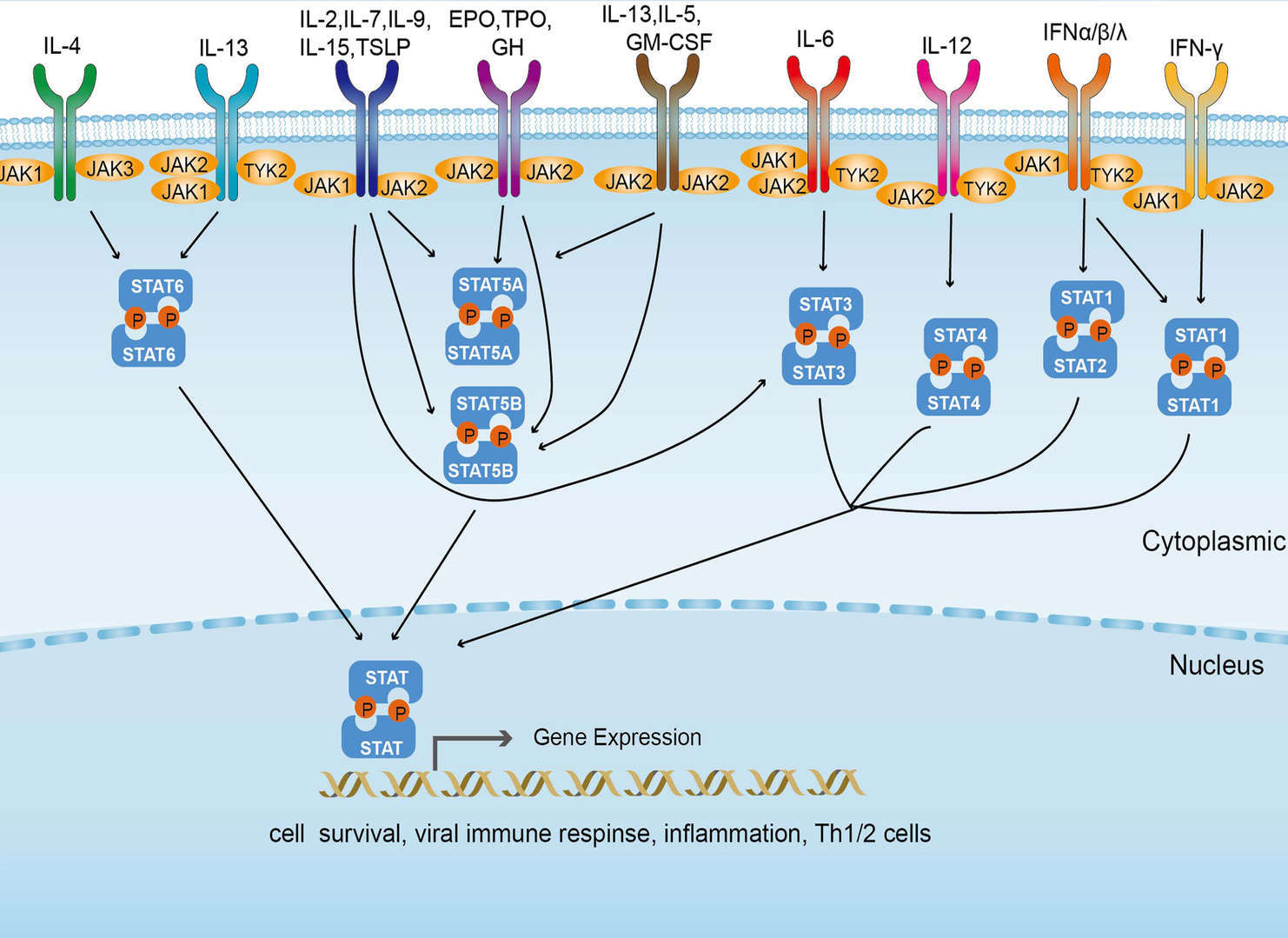 JAK-STAT Signaling Pathway
Downloadable Resources
Download resources about recombinant antibody development and antibody engineering to boost your research.
Product Notes
This is a product of Creative Biolabs' Hi-Affi™ recombinant antibody portfolio, which has several benefits including:
• Increased sensitivity
• Confirmed specificity
• High repeatability
• Excellent batch-to-batch consistency
• Sustainable supply
• Animal-free production
See more details about Hi-Affi™ recombinant antibody benefits.
Datasheet
MSDS
COA
Certificate of Analysis LookupTo download a Certificate of Analysis, please enter a lot number in the search box below. Note: Certificate of Analysis not available for kit components.
Lot Number:
Protocol & Troubleshooting
We have outlined the assay protocols, covering reagents, solutions, procedures, and troubleshooting tips for common issues in order to better assist clients in conducting experiments with our products. View the full list of Protocol & Troubleshooting.
See other products for "Cetuximab"
Afuco™ Anti-EGFR ADCC Recombinant Antibody, ADCC Enhanced (AFC-TAB-003)This product is an ADCC enhanced antibody produced by our Afuco™ platform. Recombinant monoclonal antibody to Human EGFR. It is an epidermal growth factor receptor (EGFR) inhibitor used for the treatment of metastatic colorectal cancer and head and neck cancer. It is a chimeric (mouse/human) monoclonal antibody given by intravenous infusion.
See other products for "EGFR"
Select a product category from the dropdown menu below to view related products.
| CAT | Product Name | Application | Type |
|---|---|---|---|
| TAB-750 | Anti-EGFR/HER1 Recombinant Antibody (TAB-750) | Neut, ELISA, IF, IP, FuncS, FC, WB | IgG1 - kappa |
| CAT | Product Name | Application | Type |
|---|---|---|---|
| MOB-1078z | Mouse Anti-EGFR Recombinant Antibody (clone 42C11) | WB, ELISA, FC, IF, IHC, FuncS | Mouse IgG1, κ |
| CAT | Product Name | Application | Type |
|---|---|---|---|
| NABG-056 | Recombinant Anti-Mouse Egfr VHH Single Domain Antibody | ELISA, IHC, FC, FuncS | Llama VHH |
| CAT | Product Name | Application | Type |
|---|---|---|---|
| TAB-H35 | Anti-Human EGFR Recombinant Antibody (Futuximab) | IF, WB, Inhib | IgG1 - kappa |
| CAT | Product Name | Application | Type |
|---|---|---|---|
| TAB-020 | Anti-Human EGFR Recombinant Antibody (Panitumumab) | ELISA, IP, FC, FuncS, Neut, IF, ICC | IgG2 - kappa |
| CAT | Product Name | Application | Type |
|---|---|---|---|
| TAB-165 | Anti-Human EGFR Recombinant Antibody (Matuzumab) | Neut, ELISA, IF, IP, FuncS, FC, ICC | IgG1 |
| CAT | Product Name | Application | Type |
|---|---|---|---|
| TAB-710 | Anti-EGFR Recombinant Antibody (Nimotuzumab) | ELISA, IP, FC, FuncS, Neut, IF, IHC | IgG1 - kappa |
| CAT | Product Name | Application | Type |
|---|---|---|---|
| TAB-040 | Anti-Human EGFR Recombinant Antibody (TAB-040) | ELISA, FC, IP, FuncS, IF, Neut, ICC | IgG1 - kappa |
| CAT | Product Name | Application | Type |
|---|---|---|---|
| TAB-119 | Anti-Human EGFR Recombinant Antibody (TAB-119) | FC, IP, ELISA, Neut, FuncS, IF, WB | IgG1 - kappa |
| CAT | Product Name | Application | Type |
|---|---|---|---|
| TAB-753 | Anti-EGFR Recombinant Antibody (Imgatuzumab) | Neut, ELISA, IF, IP, FuncS, FC, WB | IgG1 - kappa |
| CAT | Product Name | Application | Type |
|---|---|---|---|
| TAB-H49 | Anti-Human EGFR Recombinant Antibody (Modotuximab) | FuncS, IF, Neut, ELISA, FC, IP, IHC | IgG1 - kappa |
| CAT | Product Name | Application | Type |
|---|---|---|---|
| TAB-228CL | Anti-Human EGFR Recombinant Antibody (ABT-806) | WB, IHC | Antibody |
| CAT | Product Name | Application | Type |
|---|---|---|---|
| MOB-0242MC | Rabbit Anti-Human EGFR (phospho Y1092) Antibody | IHC, WB |
| CAT | Product Name | Application | Type |
|---|---|---|---|
| MOB-0243MC | Rabbit Anti-Human EGFR (phospho Y1068) Antibody | IHC, WB |
| CAT | Product Name | Application | Type |
|---|---|---|---|
| PABL-080 | Human Anti-EGFR Recombinant Antibody (PABL-080) | ELISA, WB, FuncS | Human IgG |
| CAT | Product Name | Application | Type |
|---|---|---|---|
| PSBL-080 | Human Anti-EGFR Recombinant Antibody; scFv Fragment (PSBL-080) | ELISA, WB, FuncS | Human scFv |
| CAT | Product Name | Application | Type |
|---|---|---|---|
| PFBL-080 | Human Anti-EGFR Recombinant Antibody; Fab Fragment (PFBL-080) | ELISA, WB, FuncS | Human Fab |
| CAT | Product Name | Application | Type |
|---|---|---|---|
| PNBL-016 | Recombinant Anti-Human EGFR VHH Single Domain Antibody (PNBL-016) | WB, ELISA | Llama VHH |
| CAT | Product Name | Application | Type |
|---|---|---|---|
| PNBL-017 | Recombinant Anti-Human EGFR VHH Single Domain Antibody (PNBL-017) | FuncS, ELISA, IF | Llama VHH |
| CAT | Product Name | Application | Type |
|---|---|---|---|
| PNBL-018 | Recombinant Anti-Human EGFR VHH Single Domain Antibody (PNBL-018) | FuncS, SPR | Llama VHH |
| CAT | Product Name | Application | Type |
|---|---|---|---|
| PABZ-039 | Mouse Anti-EGFR Recombinant Antibody (clone mAb528) | FC | Mouse IgG |
| CAT | Product Name | Application | Type |
|---|---|---|---|
| PFBZ-039 | Mouse Anti-EGFR Recombinant Antibody (clone mAb528); Fab Fragment | FC | Mouse Fab |
| CAT | Product Name | Application | Type |
|---|---|---|---|
| PFBW-039 | Human Anti-EGFR Recombinant Antibody Fab Fragment (PFBW-039) | FuncS | Chimeric (mouse/human) Fab |
| CAT | Product Name | Application | Type |
|---|---|---|---|
| PFBC-040 | Human Anti-EGFR Recombinant Antibody (clone MR1); Fab Fragment | Block | Human Fab |
| CAT | Product Name | Application | Type |
|---|---|---|---|
| PFBL-459 | Human Anti-EGFR Recombinant Antibody (clone C225); Fab Fragment | FC | Human Fab |
| CAT | Product Name | Application | Type |
|---|---|---|---|
| PFBW-171 | Mouse Anti-EGFR Recombinant Antibody; Fab Fragment (PFBW-171) | WB | Mouse Fab |
| CAT | Product Name | Application | Type |
|---|---|---|---|
| PSBZ-039 | Mouse Anti-EGFR Recombinant Antibody (clone mAb528); scFv Fragment | FC | Mouse scFv |
| CAT | Product Name | Application | Type |
|---|---|---|---|
| PSBW-039 | Mouse Anti-EGFR Recombinant Antibody scFv Fragment (PSBW-039) | Block | Mouse scFv |
| CAT | Product Name | Application | Type |
|---|---|---|---|
| PSBC-040 | Human Anti-EGFR Recombinant Antibody (clone MR1); scFv Fragment | Block | Human scFv |
| CAT | Product Name | Application | Type |
|---|---|---|---|
| TAB-0225CL | Human Anti-EGFR Recombinant Antibody (TAB-0225CL) | Block, Inhib, FuncS, Apop, In vivo | Chimeric (Mouse/Human) IgG1 |
| CAT | Product Name | Application | Type |
|---|---|---|---|
| TAB-0564CL | Mouse Anti-EGFR Recombinant Antibody (TAB-0564CL) | ELISA | Mouse IgG |
| CAT | Product Name | Application | Type |
|---|---|---|---|
| TAB-0565CL | Mouse Anti-EGFR Recombinant Antibody (TAB-0565CL) | ELISA | Mouse IgG |
| CAT | Product Name | Application | Type |
|---|---|---|---|
| TAB-0225CL-S(P) | Human Anti-EGFR Recombinant Antibody; scFv Fragment (TAB-0225CL-S(P)) | Block, Inhib, FuncS, Apop, In vivo | Human scFv |
| CAT | Product Name | Application | Type |
|---|---|---|---|
| TAB-0564CL-S(P) | Mouse Anti-EGFR Recombinant Antibody; scFv Fragment (TAB-0564CL-S(P)) | ELISA | Mouse scFv |
| CAT | Product Name | Application | Type |
|---|---|---|---|
| TAB-0565CL-S(P) | Mouse Anti-EGFR Recombinant Antibody; scFv Fragment (TAB-0565CL-S(P)) | ELISA | Mouse scFv |
| CAT | Product Name | Application | Type |
|---|---|---|---|
| TAB-0564CL-F(E) | Mouse Anti-EGFR Recombinant Antibody; Fab Fragment (TAB-0564CL-F(E)) | ELISA | Mouse Fab |
| CAT | Product Name | Application | Type |
|---|---|---|---|
| TAB-270MZ | Human Anti-EGFR Recombinant Antibody (TAB-270MZ) | ELISA | Human antibody |
| CAT | Product Name | Application | Type |
|---|---|---|---|
| TAB-274MZ | Human Anti-EGFR Recombinant Antibody (TAB-274MZ) | FC | Humanized IgG |
| CAT | Product Name | Application | Type |
|---|---|---|---|
| TAB-278MZ | Human Anti-EGFR Recombinant Antibody (TAB-278MZ) | Cyt, ELISA, Inhib | Human IgG |
| CAT | Product Name | Application | Type |
|---|---|---|---|
| TAB-015MZ-VHH | Anti-Human EGFR Recombinant Antibody (TAB-015MZ-VHH) | sELISA | Single domain antibody |
| CAT | Product Name | Application | Type |
|---|---|---|---|
| Gly-055LC | Recombinant Anti-Human EGFR Antibody (Fc glycosylation/High-mannose glycosylated) | ELISA | Chimeric antibody (mouse/human) |
| Gly-055LC-1 | Recombinant Anti-Human EGFR Antibody (Fc glycosylation/High-mannose glycosylated) | ELISA | Chimeric antibody (mouse/human) |
| CAT | Product Name | Application | Type |
|---|---|---|---|
| Gly-144LC | Recombinant Anti-Human EGFR Antibody (Fc glycosylation) | ELISA | Humanized antibody |
| CAT | Product Name | Application | Type |
|---|---|---|---|
| Gly-167LC | Recombinant Anti-Human EGFR Antibody (Non-glycosylated) | ELISA | Human antibody |
| CAT | Product Name | Application | Type |
|---|---|---|---|
| BRD-0183MZ | Chicken Anti-EGFR Polyclonal IgY | WB | Chicken antibody |
| CAT | Product Name | Application | Type |
|---|---|---|---|
| MHC-LC773 | A*0201/Human EGFR (YLNTVQPTCV) MHC Tetramer | FCM |
| CAT | Product Name | Application | Type |
|---|---|---|---|
| NEUT-722CQ | Rabbit Anti-EGFR Recombinant Antibody (clone CBL1011) | Neut | Rabbit IgG |
| CAT | Product Name | Application | Type |
|---|---|---|---|
| NEUT-723CQ | Mouse Anti-EGFR Recombinant Antibody (clone CBL931) | WB, IP, IHC, ICC, Neut | Mouse IgG1 |
| CAT | Product Name | Application | Type |
|---|---|---|---|
| NEUT-724CQ | Rabbit Anti-EGFR Recombinant Antibody (NEUT-724CQ) | IF, FC, WB, IP, Neut | Rabbit IgG |
| CAT | Product Name | Application | Type |
|---|---|---|---|
| MOR-1101 | Hi-Affi™ Rabbit Anti-EGFR Recombinant Antibody (clone DS1101AB) | IHC-P | Rabbit IgG |
| CAT | Product Name | Application | Type |
|---|---|---|---|
| MOR-4520 | Hi-Affi™ Rabbit Anti-EGFR Recombinant Antibody (clone TH28DS) | IF, ICC, FC | Rabbit IgG |
| CAT | Product Name | Application | Type |
|---|---|---|---|
| MOR-4570 | Hi-Affi™ Rabbit Anti-EGFR Recombinant Antibody (clone TH82DS) | ELISA | Rabbit IgG |
| CAT | Product Name | Application | Type |
|---|---|---|---|
| MOR-4571 | Hi-Affi™ Rabbit Anti-EGFR Recombinant Antibody (clone TH83DS) | WB, IF, ICC, FC | Rabbit IgG |
| CAT | Product Name | Application | Type |
|---|---|---|---|
| MOR-4675 | Hi-Affi™ Rabbit Anti-EGFR Recombinant Antibody (clone TH189DS) | WB, IF, ICC, FC | Rabbit IgG |
| CAT | Product Name | Application | Type |
|---|---|---|---|
| MHC-LC4545 | PE-DQB1*03:02/Human EGFR (SRALEEKKGNYVVTHG) MHC Tetramer | FCM |
| CAT | Product Name | Application | Type |
|---|---|---|---|
| AFC-TAB-165 | Afuco™ Anti-EGFR ADCC Recombinant Antibody, ADCC Enhanced (AFC-TAB-165) | Neut, ELISA, IF, IP, FuncS, FC | ADCC enhanced antibody |
| CAT | Product Name | Application | Type |
|---|---|---|---|
| AFC-TAB-464CQ | Afuco™ Anti-EGFR ADCC Recombinant Antibody, ADCC Enhanced (AFC-TAB-464CQ) | ELISA, IHC, FC, IP, IF, FuncS | ADCC enhanced antibody |
| CAT | Product Name | Application | Type |
|---|---|---|---|
| AFC-TAB-003 | Afuco™ Anti-EGFR ADCC Recombinant Antibody, ADCC Enhanced (AFC-TAB-003) | IF, IP, Neut, FuncS, ELISA, FC | ADCC enhanced antibody |
| CAT | Product Name | Application | Type |
|---|---|---|---|
| AFC-TAB-040 | Afuco™ Anti-EGFR ADCC Recombinant Antibody, ADCC Enhanced (AFC-TAB-040) | ELISA, FC, IP, FuncS, IF, Neut | ADCC enhanced antibody |
| CAT | Product Name | Application | Type |
|---|---|---|---|
| AFC-TAB-119 | Afuco™ Anti-EGFR ADCC Recombinant Antibody, ADCC Enhanced (AFC-TAB-119) | FC, IP, ELISA, Neut, FuncS, IF | ADCC enhanced antibody |
| CAT | Product Name | Application | Type |
|---|---|---|---|
| VS-0424-XY84 | AbPlus™ Anti-EGFR Magnetic Beads (pSEX81-6) | IP, Protein Purification |
| CAT | Product Name | Application | Type |
|---|---|---|---|
| VS-0924-YC32 | Mouse Anti-EGFR Recombinant Antibody (VS-0924-YC32) - Cancer Stem Cell Marker | IHC, WB | Mouse IgG1 |
| CAT | Product Name | Application | Type |
|---|---|---|---|
| VS-0924-YC35 | Rabbit Anti-EGFR Antibody (VS-0924-YC35) - Cancer Stem Cell Marker | IHC, WB, IF | Rabbit IgG |
| CAT | Product Name | Application | Type |
|---|---|---|---|
| VS-1024-XY177 | Mouse Anti-NHP EGFR Recombinant Antibody (clone 225) | IF, IP | Mouse IgG1 |
| CAT | Product Name | Application | Type |
|---|---|---|---|
| VS-0125-FY28 | Human Anti-EGFR (clone ABT-806) scFv-Fc Chimera | FC, Cyt | Human IgG1, scFv-Fc |
| CAT | Product Name | Application | Type |
|---|---|---|---|
| VS-0225-XY102 | CytoStream™ Mouse Anti-EGFR Recombinant Antibody (VS-0225-XY102) | FC | Mouse IgG1, kappa |
| CAT | Product Name | Application | Type |
|---|---|---|---|
| VS-0325-XY735 | Anti-EGFR Immunohistochemistry Kit | IHC |
| CAT | Product Name | Application | Type |
|---|---|---|---|
| VS-0425-YC340 | Recombinant Anti-EGFR Vesicular Antibody, EV Displayed (VS-0425-YC340) | ELISA, FC, Neut, Cell-uptake |
| CAT | Product Name | Application | Type |
|---|---|---|---|
| VS-0525-XY2183 | Anti-Mouse EGFR Immunohistochemistry Kit | IHC |
| CAT | Product Name | Application | Type |
|---|---|---|---|
| VS-0525-YC65 | Recombinant Anti-EGFR (AA 269-278 x AA 526-535) Biparatopic Antibody, Tandem scFv (Clone Pep 2 x Clone Pep 3) | FC | Tandem scFv |
| CAT | Product Name | Application | Type |
|---|---|---|---|
| VS-0525-YC66 | Recombinant Anti-EGFR (AA 582-591 x AA 606-614) Biparatopic Antibody, Tandem scFv (Clone Pep 4 x Clone Pep 1) | FC | Tandem scFv |
| CAT | Product Name | Application | Type |
|---|---|---|---|
| VS-0525-YC68 | Recombinant Anti-EGFR (AA 526-535 x AA 600-605) Biparatopic Antibody, Tandem scFv (Clone Pep 3 x Clone Pep 5) | FC | Tandem scFv |
| CAT | Product Name | Application | Type |
|---|---|---|---|
| VS-0525-YC213 | Recombinant Anti-EGFR (Domain II x Domain III) Biparatopic Antibody, Tandem scFv | ELISA, FC, IF, IHC, IP | Tandem scFv |
| CAT | Product Name | Application | Type |
|---|---|---|---|
| VS-0525-XY2182 | Anti-Human EGFR Immunohistochemistry Kit | IHC |
| CAT | Product Name | Application | Type |
|---|---|---|---|
| VS-0825-YC110 | SmartAb™ Recombinant Anti-EGFR pH-dependent Antibody (VS-0825-YC110) | Neut, ELISA, IF, IP, FC, WB | Human IgG1 kappa |
| CAT | Product Name | Application | Type |
|---|---|---|---|
| VS-1025-YC4 | Anti-EGFR Antibody Prodrug, Protease Activated (clone 528) | ISZ, Cyt, FuncS |
| CAT | Product Name | Application | Type |
|---|---|---|---|
| VS-1025-YC5 | Anti-EGFR Antibody Prodrug, Protease Activated (Cetuximab) | ISZ, Cyt, FuncS |
| CAT | Product Name | Application | Type |
|---|---|---|---|
| VS-1025-YC6 | Anti-EGFR Antibody Prodrug, Protease Activated (Panitumumab) | ISZ, Cyt, FuncS |
| CAT | Product Name | Application | Type |
|---|---|---|---|
| VS-1125-XY308 | Rabbit Anti-EGFR Recombinant Antibody (VS-1125-XY308) | WB, IHC, ICC, IF, FC, IP | Rabbit IgG |
| CAT | Product Name | Application | Type |
|---|---|---|---|
| VS-1125-XY309 | Rabbit Anti-EGFR Recombinant Antibody (VS-1125-XY309) | WB, ICC, IF, FC, IP | Rabbit IgG |
| CAT | Product Name | Application | Type |
|---|---|---|---|
| VS-1125-XY310 | Mouse Anti-EGFR Recombinant Antibody (VS-1125-XY310) | WB, IHC, ICC, IF, FC | Mouse IgG2a |
| CAT | Product Name | Application | Type |
|---|---|---|---|
| VS-1125-XY311 | Mouse Anti-EGFR Recombinant Antibody (VS-1125-XY311) | WB | Mouse IgG |
| CAT | Product Name | Application | Type |
|---|---|---|---|
| VS-1125-XY312 | Mouse Anti-EGFR Recombinant Antibody (VS-1125-XY312) | WB, IHC, ICC, IF, ELISA, IP | Mouse IgG1 |
Specific Inquiry

See Our Custom Production in Action


Popular Products

Application: ELISA, IP, FC, FuncS, Neut, IF, ICC

Application: WB, IF, IP, Neut, FuncS, ELISA, FC

Application: Neut, ELISA, IF, IP, FuncS, FC, ICC

Application: Neut, ELISA, IF, IP, FuncS, FC, ICC

Application: Neut, ELISA, IF, IP, FuncS, FC, ICC

Application: WB, FC, IP, ELISA, Neut, FuncS, IF

Application: Neut, ELISA, IF, IP, FuncS, FC, ICC
-2.png)
Application: ELISA, FC, IP, FuncS, IF, Neut, ICC

Application: ELISA, FC, IP, FuncS, IF, Neut, ICC

Application: FuncS, IF, Neut, ELISA, FC, IP, ICC
-2-1.png)
Application: IP, IF, FuncS, FC, Neut, ELISA, IHC


Application: ELISA, Neut, FuncS


Application: ELISA, SPR, Inhib, FuncS

For research use only. Not intended for any clinical use. No products from Creative Biolabs may be resold, modified for resale or used to manufacture commercial products without prior written approval from Creative Biolabs.
Send Inquiry
This site is protected by reCAPTCHA and the Google Privacy Policy and Terms of Service apply.