

Mouse Anti-TGFBR1 Recombinant Antibody (VS3-CJ919)
CAT#: VS3-CJ919
This product is a mouse antibody that recognizes human TGFBR1.
Gene Expression

Subcellular Location
Figure 1 IF staining of human cell line A549
Immunofluorescent staining of human cell line A549 shows localization to plasma membrane.
* Image credit: Image credit: Human Protein Atlas v21.proteinatlas.org/images/56473/985_F7_2_selected.jpg

Normal Tissue
Figure 2 IHC staining of human tonsil
Immunohistochemical staining of human tonsil shows distinct cytoplasmic positivity in reaction center cells and lymphoid cells outside reaction centra.
* Image credit: Image credit: Human Protein Atlas v21.proteinatlas.org/images/2441/ihc_selected.jpg

Normal Tissue
Figure 3 Cerebral cortex
Glial cells Staining: Low Intensity: Weak Quantity: 75%-25% Location: Cytoplasmic/ membranous Neuronal cells Staining: Low Intensity: Weak Quantity: 75%-25% Location: Cytoplasmic/ membranous Neuropil Staining: High Intensity: Strong Quantity:>75% Location: Cytoplasmic/ membranous
* Image credit: Image credit: Human Protein Atlas v21.proteinatlas.org/images/31481/66574_B_9_5.jpg

Normal Tissue
Figure 4 Cerebellum
Cells in granular layer Staining: High Intensity: Strong Quantity: 75%-25% Location: Cytoplasmic/ membranous Purkinje cells Staining: Low Intensity: Weak Quantity: 75%-25% Location: Cytoplasmic/ membranous
* Image credit: Image credit: Human Protein Atlas v21.proteinatlas.org/images/31481/66574_B_8_8.jpg

Normal Tissue
Figure 5 Colon
Endothelial cells Staining: Low Intensity: Weak Quantity:>75% Location: Cytoplasmic/ membranous Glandular cells Staining: High Intensity: Strong Quantity:>75% Location: Cytoplasmic/ membranous Peripheral nerve/ganglion Staining: Low Intensity: Weak Quantity:>75% Location: Cytoplasmic/ membranous
* Image credit: Image credit: Human Protein Atlas v21.proteinatlas.org/images/31481/66574_A_7_3.jpg

Normal Tissue
Figure 6 Kidney
Cells in tubules Staining: Medium Intensity: Moderate Quantity:>75% Location: Cytoplasmic/ membranous
* Image credit: Image credit: Human Protein Atlas v21.proteinatlas.org/images/31481/66574_A_8_5.jpg

RNA Expression
Figure 7 RNA cell line category: Cell line enhanced (RH-30)
Cell lines ordered by descending RNA expression order
* Image credit: Image credit: Human Protein Atlas v21.proteinatlas.org/ENSG00000106799-TGFBR1
❮


 ❯
❯
Specifications
- Immunogen
- KLH-conjugated synthetic peptide encompassing a sequence within human ALK5
- Host Species
- Mouse
- Type
- Mouse IgG2a
- Specificity
- Human TGFBR1
- Species Reactivity
- Human
- Applications
- IHC
- Conjugate
- Unconjugated
Product Property
- Purification
- Immunogen affinity chromatography
- Purity
- >95% as determined by SDS-PAGE
- Format
- Liquid
- Buffer
- 50% Glycerol, 0.2% BSA, PBS.
- Preservative
- 0.01% Sodium Azide
- Storage
- Store at 4°C for short term. Aliquot and store at -20°C for long term. Avoid repeated freeze/thaw cycles.
- Shipping
- Shipped at 4°C
Applications
- Application Notes
- This antibody has been tested for use in Immunohistochemistry (1:100-1:300).
Target
- Alternative Names
- AAT5; ALK5; ESS1; LDS1; MSSE; SKR4; TBRI; ALK-5; LDS1A; LDS2A; TBR-i; TGFR-1; ACVRLK4; tbetaR-I
- Gene ID
- 7046
- UniProt ID
- P36897
- Sequence Similarities
- Belongs to the protein kinase superfamily. TKL Ser/Thr protein kinase family. TGFB receptor subfamily.
- Cellular Localization
- Cell junction, Cell membrane, Membrane, Tight junction
- Post Translation Modifications
- Phosphorylated at basal levels in the absence of ligand. Activated upon phosphorylation by TGFBR2, mainly in the GS domain. Phosphorylation in the GS domain abrogates FKBP1A-binding.
N-Glycosylated.
Ubiquitinated; undergoes ubiquitination catalyzed by several E3 ubiquitin ligases including SMURF1, SMURF2 and NEDD4L2. Results in the proteasomal and/or lysosomal degradation of the receptor thereby negatively regulating its activity. Deubiquitinated by USP15, leading to stabilization of the protein and enhanced TGF-beta signal. Its ubiquitination and proteasome-mediated degradation is negatively regulated by SDCBP (PubMed:25893292).
- Protein Refseq
- NP_001124388.1; NP_001293139.1; NP_004603.1
- Function
- Transmembrane serine/threonine kinase forming with the TGF-beta type II serine/threonine kinase receptor, TGFBR2, the non-promiscuous receptor for the TGF-beta cytokines TGFB1, TGFB2 and TGFB3. Transduces the TGFB1, TGFB2 and TGFB3 signal from the cell surface to the cytoplasm and is thus regulating a plethora of physiological and pathological processes including cell cycle arrest in epithelial and hematopoietic cells, control of mesenchymal cell proliferation and differentiation, wound healing, extracellular matrix production, immunosuppression and carcinogenesis. The formation of the receptor complex composed of 2 TGFBR1 and 2 TGFBR2 molecules symmetrically bound to the cytokine dimer results in the phosphorylation and the activation of TGFBR1 by the constitutively active TGFBR2. Activated TGFBR1 phosphorylates SMAD2 which dissociates from the receptor and interacts with SMAD4. The SMAD2-SMAD4 complex is subsequently translocated to the nucleus where it modulates the transcription of the TGF-beta-regulated genes. This constitutes the canonical SMAD-dependent TGF-beta signaling cascade. Also involved in non-canonical, SMAD-independent TGF-beta signaling pathways. For instance, TGFBR1 induces TRAF6 autoubiquitination which in turn results in MAP3K7 ubiquitination and activation to trigger apoptosis. Also regulates epithelial to mesenchymal transition through a SMAD-independent signaling pathway through PARD6A phosphorylation and activation.
REVIEWS AND Q&AS
CITATIONS
RESOURCES
DOWNLOADS
RELATED PRODUCTS
Inquiry
Navs
Customer Review

There are currently no Customer reviews or questions for VS3-CJ919. Click the button above to contact us or submit your feedback about this product.
Submit Your Publication
Published with our product? Submit your paper and receive a 10% discount on your next order! Share your research to earn exclusive rewards.
Related Signaling Pathways
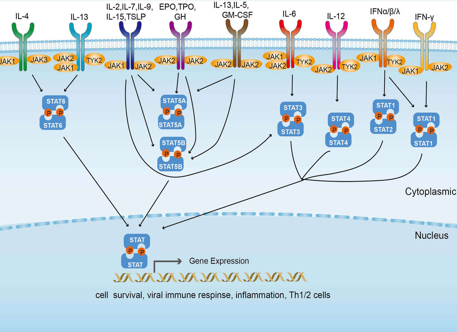 JAK-STAT Signaling Pathway
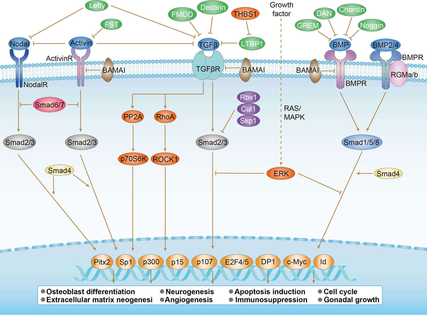 TGF-β Signaling Pathway
Related Diseases
 Pancreatic Cancer
Downloadable Resources
Download resources about recombinant antibody development and antibody engineering to boost your research.
Product Notes
This is a product of Creative Biolabs' Hi-Affi™ recombinant antibody portfolio, which has several benefits including:
• Increased sensitivity
• Confirmed specificity
• High repeatability
• Excellent batch-to-batch consistency
• Sustainable supply
• Animal-free production
See more details about Hi-Affi™ recombinant antibody benefits.
Datasheet
MSDS
COA
Certificate of Analysis LookupTo download a Certificate of Analysis, please enter a lot number in the search box below. Note: Certificate of Analysis not available for kit components.
Lot Number:
Protocol & Troubleshooting
We have outlined the assay protocols, covering reagents, solutions, procedures, and troubleshooting tips for common issues in order to better assist clients in conducting experiments with our products. View the full list of Protocol & Troubleshooting.
Secondary Antibody
- CAT
- Product Name
Recommended Dilution Buffer
- CAT
- Product Name
See other products for "TGFBR1"
Select a product category from the dropdown menu below to view related products.
| CAT | Product Name | Application | Type |
|---|---|---|---|
| MOB-4032z | Mouse Anti-TGFBR1 Recombinant Antibody (clone 43H12) | WB, ELISA, IHC | Mouse IgG1 |
| CAT | Product Name | Application | Type |
|---|---|---|---|
| AGTO-L077E | TGFα-PE immunotoxin | Cytotoxicity assay, Functional assay |
| CAT | Product Name | Application | Type |
|---|---|---|---|
| MOB-2021CT | Recombinant Mouse anti-Human TGFBR1 Monoclonal antibody (9B22) | IHC-P |
| CAT | Product Name | Application | Type |
|---|---|---|---|
| ZG-0114C | Mouse Anti-TGFBR1 Recombinant Antibody (clone ABT-TGFR1) | IHC-P, IF | Mouse IgG |
| CAT | Product Name | Application | Type |
|---|---|---|---|
| ZG-0115C | Mouse Anti-TGFBR1 Recombinant Antibody (clone ABT-TGFR2) | IHC-P, IF | Mouse IgG |
| CAT | Product Name | Application | Type |
|---|---|---|---|
| VS3-FY1446 | Recombinant Mouse Anti-TGFBR1 Antibody (clone 4H5-6E6-7H2) | IHC-P | Mouse IgG2a |
| CAT | Product Name | Application | Type |
|---|---|---|---|
| VS-0425-YC39 | Recombinant Anti-Tgfbr1 Vesicular Antibody, EV Displayed (VS-0425-YC39) | ELISA, FC, Cell-uptake |
| CAT | Product Name | Application | Type |
|---|---|---|---|
| VS-0525-XY7216 | Anti-Mouse TGFBR1 Immunohistochemistry Kit | IHC |
Specific Inquiry

See Our Custom Production in Action


Popular Products

Application: Neut, ELISA, IF, IP, FuncS, FC, ICC

Application: IF, IP, Neut, FuncS, ELISA, FC, ICC

Application: IF, IP, Neut, FuncS, ELISA, FC, WB

Application: ELISA, FC, IP, FuncS, IF, Neut, ICC
-2.png)
Application: FuncS, IF, Neut, ELISA, FC, IP, ICC

Application: WB, FuncS, IF, Neut, ELISA, FC, IP


Application: WB, IP, IF, FuncS, FC, Neut, ELISA


Application: ELISA, FC, IP, FuncS, IF, Neut, ICC

Application: FuncS, IF, Neut, ELISA, FC, IP, WB

Application: IF, IP, Neut, FuncS, ELISA, FC, ICC

Application: WB, Neut, ELISA, IF, IP, FuncS, FC


Application: ELISA, WB, BLI, SPR

Application: Block, Cyt, FuncS, Inhib


For research use only. Not intended for any clinical use. No products from Creative Biolabs may be resold, modified for resale or used to manufacture commercial products without prior written approval from Creative Biolabs.
Send Inquiry
This site is protected by reCAPTCHA and the Google Privacy Policy and Terms of Service apply.