Mouse Anti-MYC Recombinant Antibody (clone 9E10)
CAT#: PABL-281
Recombinant mouse antibody (9E10) is capable of binding to MYC. The murine monoclonal antibody 9E10, which was raised against a peptide from the C-terminal region of the human c-myc protooncogene product, is widely used in cell biology and protein engineering as a detection or purification tool for proteins that are fused with the corresponding epitope as an affinity tag.


Specifications
- Immunogen
- Human v-myc avian myelocytomatosis viral oncogene homolog
- Host Species
- Mouse
- Derivation
- Mouse
- Type
- Mouse IgG
- Specificity
- Human MYC
- Species Reactivity
- Human
- Clone
- 9E10
- Applications
- WB, IHC, FuncS
Product Property
- Purity
- >95% as determined by SDS-PAGE and HPLC analysis
- Concentration
- Please refer to the vial label for the specific concentration.
- Buffer
- PBS
- Preservative
- No preservatives
- Storage
- Centrifuge briefly prior to opening vial. Store at +4°C short term (1-2 weeks). Aliquot and store at -20°C long term. Avoid repeated freeze/thaw cycles.
Target
- Alternative Names
- MYC; v-myc avian myelocytomatosis viral oncogene homolog; MRTL; MYCC; c-Myc; bHLHe39; myc proto-oncogene protein; proto-oncogene c-Myc; transcription factor p64; class E basic helix-loop-helix protein 39; avian myelocytomatosis viral oncogene homolog; v-myc myelocytomatosis viral oncogene homolog; myc-related translation/localization regulatory factor
- Gene ID
- 4609
- UniProt ID
- P01106
Customer Review
There are currently no Customer reviews or questions for PABL-281. Click the button above to contact us or submit your feedback about this product.
Submit Your Publication
Published with our product? Submit your paper and receive a 10% discount on your next order! Share your research to earn exclusive rewards.
Related Signaling Pathways
Related Diseases
Downloadable Resources
Download resources about recombinant antibody development and antibody engineering to boost your research.
Product Notes
This is a product of Creative Biolabs' Hi-Affi™ recombinant antibody portfolio, which has several benefits including:
• Increased sensitivity
• Confirmed specificity
• High repeatability
• Excellent batch-to-batch consistency
• Sustainable supply
• Animal-free production
See more details about Hi-Affi™ recombinant antibody benefits.
Datasheet
MSDS
COA
Certificate of Analysis LookupTo download a Certificate of Analysis, please enter a lot number in the search box below. Note: Certificate of Analysis not available for kit components.
Protocol & Troubleshooting
We have outlined the assay protocols, covering reagents, solutions, procedures, and troubleshooting tips for common issues in order to better assist clients in conducting experiments with our products. View the full list of Protocol & Troubleshooting.
See other products for "Clone 9E10"
- CAT
- Product Name
See other products for "MYC"
Select a product category from the dropdown menu below to view related products.
| CAT | Product Name | Application | Type |
|---|---|---|---|
| PSBL-281 | Mouse Anti-MYC Recombinant Antibody (clone 9E10); scFv Fragment | WB, IHC, FuncS | Mouse scFv |
| CAT | Product Name | Application | Type |
|---|---|---|---|
| PFBL-281 | Mouse Anti-MYC Recombinant Antibody (clone 9E10); Fab Fragment | WB, IHC, FuncS | Mouse Fab |
| CAT | Product Name | Application | Type |
|---|---|---|---|
| MOB-0400MC | Anti-c-Myc antibody (HRP) | ELISA, IHC-P, IHC-Fr, WB |
| CAT | Product Name | Application | Type |
|---|---|---|---|
| MOB-0268CT | Recombinant Mouse anti-Human MYC Monoclonal antibody (4H40) | ELISA, ICC, IF, IHC-P, IP, WB |
| CAT | Product Name | Application | Type |
|---|---|---|---|
| BRD-0380MZ | Chicken Anti-c-Myc (ab1) Polyclonal IgY | Indirect ELISA, WB | Chicken antibody |
| CAT | Product Name | Application | Type |
|---|---|---|---|
| BRD-0381MZ | Chicken Anti-c-Myc (ab2) Polyclonal IgY | WB | Chicken antibody |
| CAT | Product Name | Application | Type |
|---|---|---|---|
| MOR-2336 | Hi-Affi™ Recombinant Rabbit Anti-MYC Monoclonal Antibody (DS2336AB) | WB, IP, ICC | IgG |
| CAT | Product Name | Application | Type |
|---|---|---|---|
| MOR-4554 | Hi-Affi™ Recombinant Rabbit Anti-MYC Monoclonal Antibody (TH64DS) | WB, IF, ICC, FC | IgG |
| CAT | Product Name | Application | Type |
|---|---|---|---|
| FAMAB-0112-CN | Mouse Anti-MYC Recombinant Antibody (clone CT14) | ELISA, WB, FC, Inhib | Mouse IgG2a |
| CAT | Product Name | Application | Type |
|---|---|---|---|
| FAMAB-0112-CN-S(P) | Mouse Anti-MYC Recombinant Antibody (clone CT14); scFv Fragment | ELISA, WB, FC | Mouse scFv |
| CAT | Product Name | Application | Type |
|---|---|---|---|
| FAMAB-0112-CN-F(E) | Mouse Anti-MYC Recombinant Antibody (clone CT14); Fab Fragment | ELISA, WB, FC | Mouse Fab |
| CAT | Product Name | Application | Type |
|---|---|---|---|
| MRO-1778-CN | Rabbit Anti-MYC Polyclonal Antibody (MRO-1778-CN) | WB | Rabbit IgG |
| CAT | Product Name | Application | Type |
|---|---|---|---|
| MRO-1779-CN | Rabbit Anti-MYC Polyclonal Antibody (MRO-1779-CN) | WB | Rabbit IgG |
| CAT | Product Name | Application | Type |
|---|---|---|---|
| MRO-2311-CN | Recombinant Rabbit Anti-MYC (phosphorylated Thr58) Monoclonal Antibody (CBACN-604) | WB, IF, FC | Rabbit IgG |
| CAT | Product Name | Application | Type |
|---|---|---|---|
| MOR-0050-FY | Rabbit Anti-MYC Recombinant Antibody (clone AFY0021) | ICC, IHC, WB | Rabbit IgG |
| CAT | Product Name | Application | Type |
|---|---|---|---|
| VS-0424-XY194 | AbPlus™ Anti-MYC Magnetic Beads (33) | IP, Protein Purification |
| CAT | Product Name | Application | Type |
|---|---|---|---|
| VS-1024-XY133 | Mouse Anti-NHP MYC Recombinant Antibody (clone MYC699) | IF, FC, ELISA | Mouse IgG1, kappa |
| CAT | Product Name | Application | Type |
|---|---|---|---|
| VS-0525-XY4607 | Anti-MYC Immunohistochemistry Kit | IHC |
| CAT | Product Name | Application | Type |
|---|---|---|---|
| VS-0525-XY4608 | Anti-Human MYC Immunohistochemistry Kit | IHC |
| CAT | Product Name | Application | Type |
|---|---|---|---|
| VS-0525-XY4609 | Anti-Mouse MYC Immunohistochemistry Kit | IHC |
| CAT | Product Name | Application | Type |
|---|---|---|---|
| VS-0825-YC263 | SmartAb™ Recombinant Anti-MYC pH-dependent Antibody (Clone CT14) | ELISA, WB, FC, Inhib | Mouse IgG2a |
| CAT | Product Name | Application | Type |
|---|---|---|---|
| VS-1025-YC55 | Anti-MYC Antibody Prodrug, Protease Activated (CT14) | ISZ, Cyt, FuncS |
Popular Products

Application: IP, IF, FuncS, FC, Neut, ELISA, IHC

Application: FuncS, IF, Neut, ELISA, FC, IP, ICC

Application: WB, FC, IP, ELISA, Neut, FuncS, IF

Application: ELISA, IP, FC, FuncS, Neut, IF, ICC

Application: IF, IP, Neut, FuncS, ELISA, FC, ICC

Application: IP, IF, FuncS, FC, Neut, ELISA, ICC

Application: FuncS, IF, Neut, ELISA, FC, IP, IHC

Application: IF, IP, Neut, FuncS, ELISA, FC, ICC

Application: Block, Cyt, FuncS, Inhib
For research use only. Not intended for any clinical use. No products from Creative Biolabs may be resold, modified for resale or used to manufacture commercial products without prior written approval from Creative Biolabs.
This site is protected by reCAPTCHA and the Google Privacy Policy and Terms of Service apply.


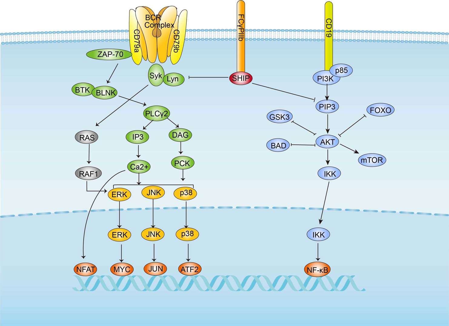 BCR Signaling Pathway
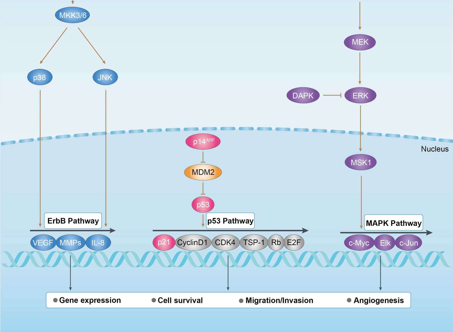 Bladder Cancer
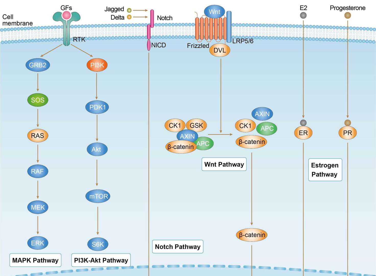 Breast Cancer
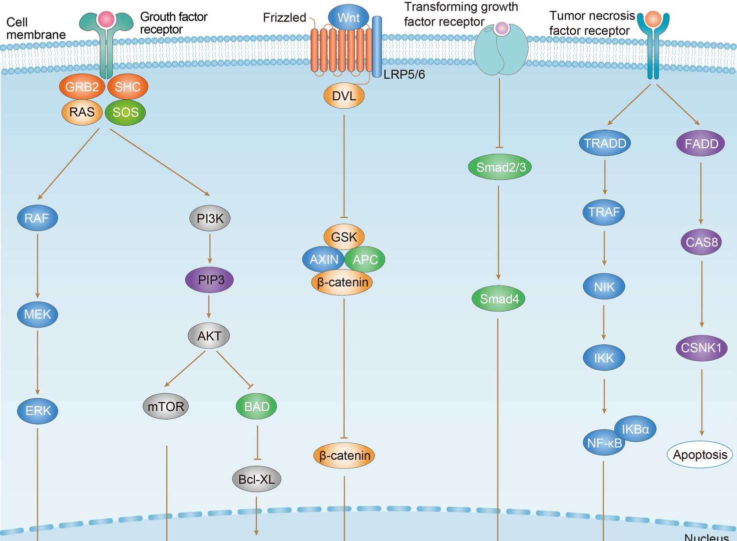 Colorectal Cancer
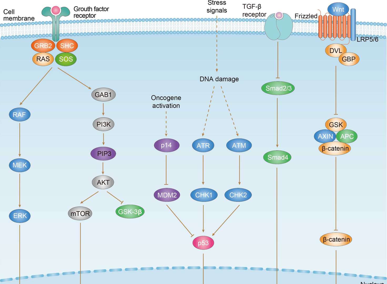 Gastric Cancer


