

Anti-CD86 (clone Hu3D1) Recombinant Antibody Coupled Liposome (VS-1024-FY6)
CAT#: VS-1024-FY6
Anti-CD86 antibody liposomes target dendritic cells (DCs) expressing CD86, providing a co-stimulatory signal for the immune response. They enhance the antigen-presenting capacity of DCs, promoting T cell activation and proliferation. The mechanism of action of these liposomes may include the enhancement of cytokine production by DCs, thereby activating downstream immune responses. Ultimately, this strategy may improve anti-tumor immunity in cancer immunotherapy.
Gene Expression

Subcellular Location
Figure 1 IF staining of human cell line HDLM-2
Immunofluorescent staining of human cell line HDLM-2 shows localization to plasma membrane & centriolar satellites.
* Image credit: Image credit: Human Protein Atlas v21.proteinatlas.org/images/26802/1962_A12_6_selected.jpg

Normal Tissue
Figure 2 Cerebral cortex
Neuronal cells Staining: Medium Intensity: Moderate Quantity: 75%-25% Location: Cytoplasmic/ membranous
* Image credit: Image credit: Human Protein Atlas v21.proteinatlas.org/images/4319/11663_B_8_5.jpg

Normal Tissue
Figure 3 Lung
Macrophages Staining: Medium Intensity: Moderate Quantity:>75% Location: Cytoplasmic/ membranous
* Image credit: Image credit: Human Protein Atlas v21.proteinatlas.org/images/4319/11663_A_1_4.jpg

Normal Tissue
Figure 4 Colon
Glandular cells Staining: Medium Intensity: Moderate Quantity: 75%-25% Location: Cytoplasmic/ membranous Peripheral nerve/ganglion Staining: Low Intensity: Weak Quantity:>75% Location: Cytoplasmic/ membranous
* Image credit: Image credit: Human Protein Atlas v21.proteinatlas.org/images/4319/11663_A_8_3.jpg

Normal Tissue
Figure 5 Lymph node
Germinal center cells Staining: High Intensity: Strong Quantity: 75%-25% Location: Cytoplasmic/ membranous Non-germinal center cells Staining: Medium Intensity: Strong Quantity: <25% Location: Cytoplasmic/ membranous
* Image credit: Image credit: Human Protein Atlas v21.proteinatlas.org/images/4319/11663_A_9_8.jpg

RNA Expression
Figure 6 RNA cell line category: Cell line enriched (HDLM-2)
Cell lines ordered by descending RNA expression order
* Image credit: Image credit: Human Protein Atlas v21.proteinatlas.org/ENSG00000114013-CD86
❮


 ❯
❯
More Infomation
Specifications
- Potential Clinical Applications
- Specific targeting of dendritic cells
Product Composition
- Clone
- Hu3D1
- Antibody Type
- IgG
- Antibody Host
- Human
- Antibody Reactivity
- Human
- Antibody Description
- The anti-B7-2 antibody is a humanized monoclonal antibody that specifically targets the B7-2 molecule found on antigen-presenting cells (APCs). This antibody has potential applications in treating autoimmune diseases, preventing transplant rejection, addressing inflammatory disorders, and managing infectious diseases.
Product Property
- Storage
- See in the COA
- Storage Shelf Time
- See in the COA
Target Information
- Target
- CD86
- Introduction
- CD86 (CD86 Molecule) is a Protein Coding gene. Diseases associated with CD86 include Gallbladder Squamous Cell Carcinoma and Myocarditis. Among its related pathways are Cytokine Signaling in Immune system and Th2 Differentiation Pathway. Gene Ontology (GO) annotations related to this gene include receptor binding and coreceptor activity. An important paralog of this gene is CD80.
- Alternative Names
- CD86 Molecule; CD86 Antigen (CD28 Antigen Ligand 2, B7-2 Antigen); CTLA-4 Counter-Receptor B7.2; CD28LG2; FUN-1; BU63; B70; B-Lymphocyte Activation Antigen B7-2
- Full Name
- CD86 Molecule
- Gene ID
- 942
- UniProt ID
- P42081
- Cellular Localization
- Cell membrane
- Post Translation Modifications
- Polyubiquitinated; which is promoted by MARCH8 and results in endocytosis and lysosomal degradation. (P42081-CD86_HUMAN)
Glycosylation at Asn135, Asn33, Asn146, Asn47, Asn154, Asn177, Asn192, and Asn213 (NX_P42081 [NX_P42081-1])
Modification sites at PhosphoSitePlus (P42081)
Glycosylation from GlyGen (P42081) 8 sites
- Protein Refseq
- NP_001193853.2; NP_001193854.2; NP_008820.4; NP_787058.5; NP_795711.2
- Function
- May play a critical role in the early events of T-cell activation and costimulation of naive T-cells, such as deciding between immunity and anergy that is made by T-cells within 24 hours after activation.
REVIEWS AND Q&AS
CITATIONS
RESOURCES
DOWNLOADS
RELATED PRODUCTS
Inquiry
Navs
Customer Review

There are currently no Customer reviews or questions for VS-1024-FY6. Click the button above to contact us or submit your feedback about this product.
Submit Your Publication
Published with our product? Submit your paper and receive a 10% discount on your next order! Share your research to earn exclusive rewards.
Related Diseases
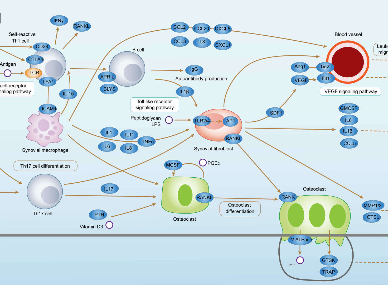 Rheumatoid Arthritis
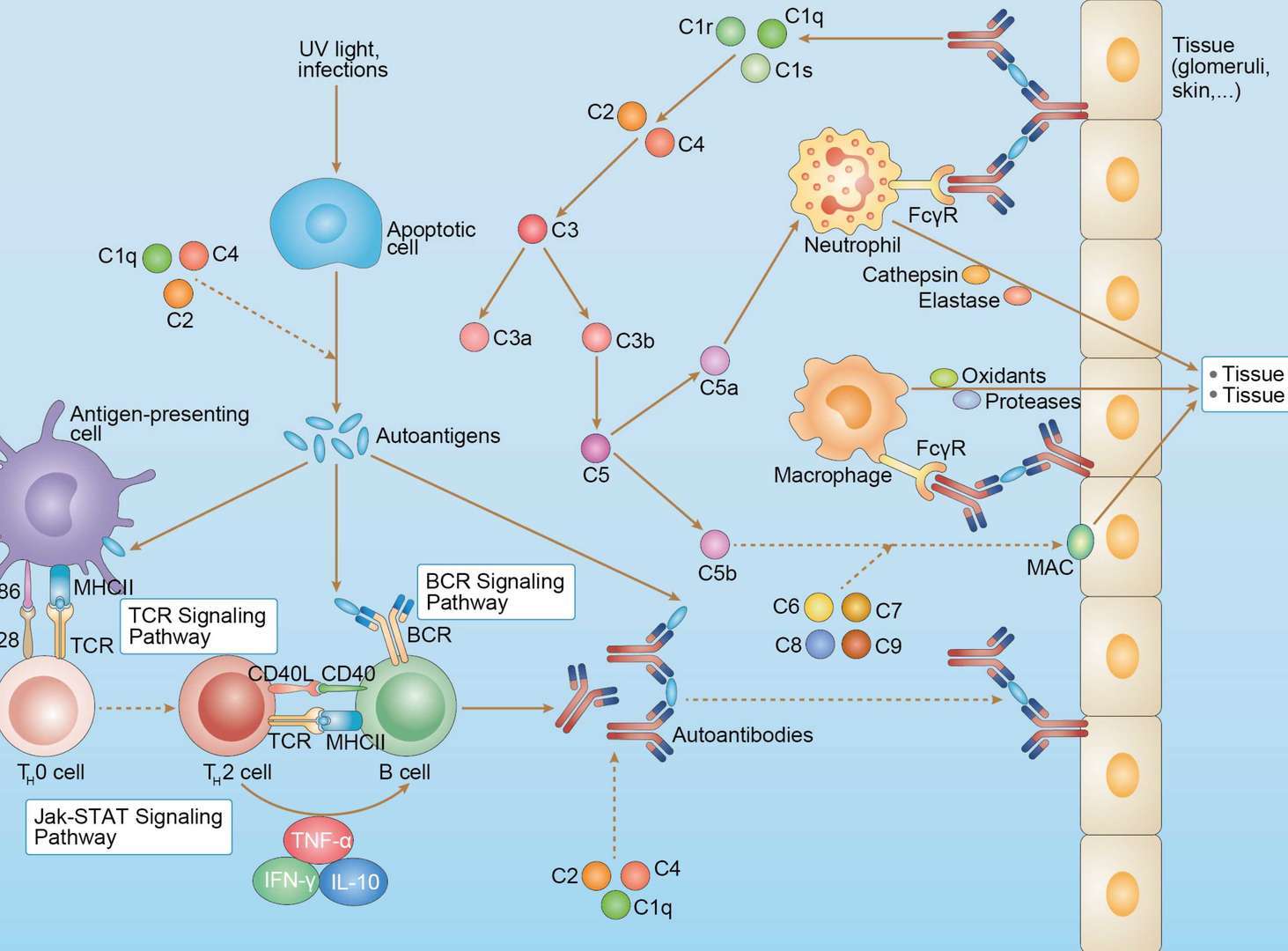 Systemic Lupus Erythematosus
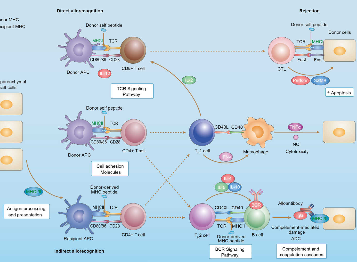 Allograft Rejection
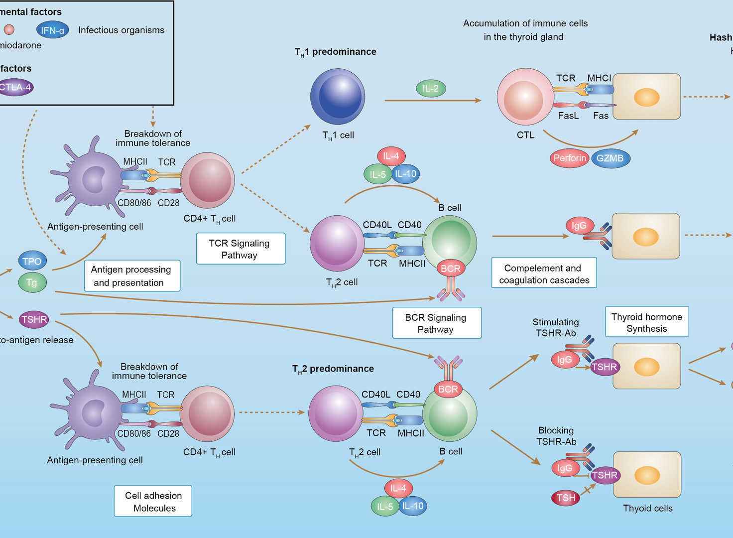 Autoimmune Thyroid Disease
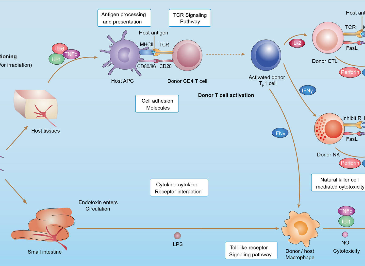 Graft-versus-Host Disease
 Maturity Onset Diabetes of the Young
Downloadable Resources
Download resources about recombinant antibody development and antibody engineering to boost your research.
Datasheet
MSDS
COA
Certificate of Analysis LookupTo download a Certificate of Analysis, please enter a lot number in the search box below. Note: Certificate of Analysis not available for kit components.
Lot Number:
See other products for "Clone Hu3D1"
- CAT
- Product Name
See other products for "CD86"
Select a product category from the dropdown menu below to view related products.
| CAT | Product Name | Application | Type |
|---|---|---|---|
| MOB-1540z | Mouse Anti-CD86 Recombinant Antibody (clone 24B7) | WB, ELISA, FC, IF, IHC, IP, FuncS | Mouse IgG1, κ |
| CAT | Product Name | Application | Type |
|---|---|---|---|
| AGTO-L002G | anti-CD86 immunotoxin 1G10 (IgG)-Gel | Cytotoxicity assay, Functional assay |
| CAT | Product Name | Application | Type |
|---|---|---|---|
| AGTO-L002S | anti-CD86 immunotoxin 1G10 (IgG)-Sap | Cytotoxicity assay, Functional assay |
| CAT | Product Name | Application | Type |
|---|---|---|---|
| AGTO-L002O | anti-CD86 immunotoxin 1G10 (IgG)-Bouganin | Cytotoxicity assay, Functional assay |
| CAT | Product Name | Application | Type |
|---|---|---|---|
| TAB-361LC | Anti-Human CD86 Recombinant Antibody (chFun1) | IHC | Chimeric antibody (mouse/human) |
| CAT | Product Name | Application | Type |
|---|---|---|---|
| TAB-361LC-S(P) | Anti-Human CD86 Recombinant Antibody scFv Fragment (chFun1) | IHC | Chimeric antibody (mouse/human) |
| CAT | Product Name | Application | Type |
|---|---|---|---|
| TAB-361LC-F(E) | Anti-Human CD86 Recombinant Antibody Fab Fragment (chFun1) | IHC | Chimeric antibody (mouse/human) |
| CAT | Product Name | Application | Type |
|---|---|---|---|
| MOB-0361MZ | Recombinant Mouse Anti-Human CD86 Molecule Antibody (clone CU64) | ELISA | Mouse antibody |
| CAT | Product Name | Application | Type |
|---|---|---|---|
| NEUT-419CQ | Mouse Anti-CD86 Recombinant Antibody (clone MM0101-7F35) | WB, Neut, FC, CyTOF® | Mouse IgG1 |
| CAT | Product Name | Application | Type |
|---|---|---|---|
| NEUT-420CQ | Mouse Anti-CD86 Recombinant Antibody (clone CBL763) | WB, FC, CyTOF®, Neut | Mouse IgG1 |
| CAT | Product Name | Application | Type |
|---|---|---|---|
| NEUT-421CQ | Mouse Anti-CD86 Recombinant Antibody (clone CBL024) | Neut, FC | Mouse IgG1 |
| CAT | Product Name | Application | Type |
|---|---|---|---|
| NEUT-422CQ | Mouse Anti-CD86 Recombinant Antibody (clone IT2.2) | FC, FuncS, Neut | Mouse IgG2b, κ |
| CAT | Product Name | Application | Type |
|---|---|---|---|
| NEUT-423CQ | Rat Anti-Cd86 Recombinant Antibody (clone 2D10), Biotin | FC, IHC-Fr, IP, Block | Rat IgG2b, κ |
| CAT | Product Name | Application | Type |
|---|---|---|---|
| NEUT-424CQ | Rat Anti-Cd86 Recombinant Antibody (clone GL-1) | FC, Block, IF, IHC, IP | Rat IgG2a, κ |
| CAT | Product Name | Application | Type |
|---|---|---|---|
| NEUT-425CQ | Rat Anti-Cd86 Recombinant Antibody (NEUT-425CQ) | Block, FC, IHC-Fr, IP | Rat IgG2 |
| CAT | Product Name | Application | Type |
|---|---|---|---|
| NEUT-426CQ | Rat Anti-Cd86 Recombinant Antibody (clone PO3.1) | FC, FuncS, Neut | Rat IgG2b, κ |
| CAT | Product Name | Application | Type |
|---|---|---|---|
| MOR-0588 | Hi-Affi™ Rabbit Anti-CD86 Recombinant Antibody (clone DS588AB) | WB, FC, IHC | Rabbit IgG |
| CAT | Product Name | Application | Type |
|---|---|---|---|
| MRO-0320-CN | Rabbit Anti-CD86 Recombinant Antibody (clone CBACN-122) | WB, IF, IHC, IP, FC | Rabbit IgG |
| CAT | Product Name | Application | Type |
|---|---|---|---|
| FN-045CQ | Mouse Anti-CD86 Recombinant Antibody (clone BU63) | FC, Block | Mouse IgG1 |
| CAT | Product Name | Application | Type |
|---|---|---|---|
| FN-096CQ | Rat Anti-CD86 Recombinant Antibody (clone GL1) | IHC, FC, Block, IF | Rat IgG2a, κ |
| CAT | Product Name | Application | Type |
|---|---|---|---|
| HPAB-1586WJ-F(E) | Mouse Anti-CD86 Recombinant Antibody; Fab Fragment (clone 3D1) | ELISA, WB | Mouse Fab |
| CAT | Product Name | Application | Type |
|---|---|---|---|
| HPAB-1586WJ-S(P) | Mouse Anti-CD86 Recombinant Antibody; scFv Fragment (clone 3D1) | ELISA, WB | Mouse scFv |
| CAT | Product Name | Application | Type |
|---|---|---|---|
| VS-0724-YC1509 | AbPlus™ Anti-Cd86 Magnetic Beads (VS-0724-YC1509) | IP, Protein Purification |
| CAT | Product Name | Application | Type |
|---|---|---|---|
| VS-0225-XY76 | CytoStream™ Mouse Anti-CD86 Recombinant Antibody (VS-0225-XY76) | FC | Mouse IgG1, kappa |
| CAT | Product Name | Application | Type |
|---|---|---|---|
| VS-0225-XY77 | CytoStream™ Mouse Anti-CD86 Recombinant Antibody (VS-0225-XY77) | FC | Mouse IgG2b, kappa |
| CAT | Product Name | Application | Type |
|---|---|---|---|
| VS13-YC179 | CytoStream™ Rat Anti-Mouse Cd86 Recombinant Antibody (VS13-YC179) | FC | Rat IgG2a |
| CAT | Product Name | Application | Type |
|---|---|---|---|
| VS-0425-FY10 | Human Anti-CD86 (clone Hu3D1) scFv-Fc Chimera | Inhib | Human IgG1, scFv-Fc |
| CAT | Product Name | Application | Type |
|---|---|---|---|
| VS-0425-YC683 | Recombinant Anti-CD86 Vesicular Antibody, EV Displayed (VS-0425-YC683) | Block, ELISA, FC, Cell-uptake |
| CAT | Product Name | Application | Type |
|---|---|---|---|
| VS-0525-XY1235 | Anti-Mouse CD86 Immunohistochemistry Kit | IHC |
| CAT | Product Name | Application | Type |
|---|---|---|---|
| VS-0525-XY1236 | Anti-Rat CD86 Immunohistochemistry Kit | IHC |
| CAT | Product Name | Application | Type |
|---|---|---|---|
| VS-0825-YC71 | SmartAb™ Recombinant Anti-CD86 pH-dependent Antibody (Clone Hu3D1) | ELISA | Human IgG2/G4 |
See Our Custom Production in Action


Popular Products

Application: WB, FC, IP, ELISA, Neut, FuncS, IF

Application: WB, ELISA, IP, FC, FuncS, Neut, IF

Application: IF, IP, Neut, FuncS, ELISA, FC, ICC

Application: ELISA, FC, IP, FuncS, IF, Neut, ICC

Application: ELISA, IP, FC, FuncS, Neut, IF, ICC

Application: ELISA, FC, IP, FuncS, IF, Neut, ICC

Application: WB, IP, IF, FuncS, FC, Neut, ELISA


Application: IP, IF, FuncS, FC, Neut, ELISA, ICC

Application: IP, IF, FuncS, FC, Neut, ELISA, ICC

Application: Neut, ELISA, IF, IP, FuncS, FC, ICC

Application: IF, IP, Neut, FuncS, ELISA, FC, ICC

Application: ELISA, FC, IP, FuncS, IF, Neut, ICC

Application: ELISA, FC, IP, FuncS, IF, Neut, ICC

Application: FuncS, IF, Neut, ELISA, FC, IP, IHC


For research use only. Not intended for any clinical use. No products from Creative Biolabs may be resold, modified for resale or used to manufacture commercial products without prior written approval from Creative Biolabs.
Send Inquiry
This site is protected by reCAPTCHA and the Google Privacy Policy and Terms of Service apply.