Rabbit Anti-MAPK1 Recombinant Antibody (VS3-CJ845)
CAT#: VS3-CJ845
This product is a rabbit antibody that recognizes human, mouse, and rat MAPK1.


Specifications
- Immunogen
- Recombinant protein
- Host Species
- Rabbit
- Type
- Rabbit IgG
- Specificity
- Human, Mouse, Rat MAPK1
- Species Reactivity
- Human, Mouse, Rat
- Applications
- WB, ICC, IF, IHC, IP, FC
- Conjugate
- Unconjugated
Product Property
- Purification
- Protein A affinity purified
- Purity
- >95% as determined by SDS-PAGE
- Format
- Liquid
- Buffer
- 40% Glycerol, 1% BSA, TBS, pH7.4.
- Preservative
- 0.05% Sodium Azide
- Storage
- Store at 4°C for short term. Aliquot and store at -20°C for long term. Avoid repeated freeze/thaw cycles.
Applications
- Application Notes
- This antibody has been tested for use in Western Blot, Immunocytochemistry, Immunofluorescence, Immunohistochemistry, Immunoprecipitation, Flow Cytometry.
Target
- Alternative Names
- ERK; p38; p40; p41; ERK2; ERT1; NS13; ERK-2; MAPK2; PRKM1; PRKM2; P42MAPK; p41mapk; p42-MAPK
- Gene ID
- 5594
- UniProt ID
- P28482
- Sequence Similarities
- Belongs to the protein kinase superfamily. CMGC Ser/Thr protein kinase family. MAP kinase subfamily.
- Cellular Localization
- Cytoplasm, Cytoskeleton, Membrane, Nucleus
- Post Translation Modifications
- Phosphorylated upon KIT and FLT3 signaling (By similarity).
Dually phosphorylated on Thr-185 and Tyr-187, which activates the enzyme. Undergoes regulatory phosphorylation on additional residues such as Ser-246 and Ser-248 in the kinase insert domain (KID) These phosphorylations, which are probably mediated by more than one kinase, are important for binding of MAPK1/ERK2 to importin-7 (IPO7) and its nuclear translocation. In addition, autophosphorylation of Thr-190 was shown to affect the subcellular localization of MAPK1/ERK2 as well. Ligand-activated ALK induces tyrosine phosphorylation. Dephosphorylated by PTPRJ at Tyr-187. Phosphorylation on Ser-29 by SGK1 results in its activation by enhancing its interaction with MAP2K1/MEK1 and MAP2K2/MEK2. DUSP3 and DUSP6 dephosphorylate specifically MAPK1/ERK2 and MAPK3/ERK1 whereas DUSP9 dephosphorylates a broader range of MAPKs. Dephosphorylated by DUSP1 and DUSP2 at Thr-185 and Tyr-187 (By similarity) (PubMed:16288922).
ISGylated.
- Protein Refseq
- NP_002736.3; NP_620407.1
- Function
- Serine/threonine kinase which acts as an essential component of the MAP kinase signal transduction pathway. MAPK1/ERK2 and MAPK3/ERK1 are the 2 MAPKs which play an important role in the MAPK/ERK cascade. They participate also in a signaling cascade initiated by activated KIT and KITLG/SCF. Depending on the cellular context, the MAPK/ERK cascade mediates diverse biological functions such as cell growth, adhesion, survival and differentiation through the regulation of transcription, translation, cytoskeletal rearrangements. The MAPK/ERK cascade plays also a role in initiation and regulation of meiosis, mitosis, and postmitotic functions in differentiated cells by phosphorylating a number of transcription factors. About 160 substrates have already been discovered for ERKs. Many of these substrates are localized in the nucleus, and seem to participate in the regulation of transcription upon stimulation. However, other substrates are found in the cytosol as well as in other cellular organelles, and those are responsible for processes such as translation, mitosis and apoptosis. Moreover, the MAPK/ERK cascade is also involved in the regulation of the endosomal dynamics, including lysosome processing and endosome cycling through the perinuclear recycling compartment (PNRC); as well as in the fragmentation of the Golgi apparatus during mitosis. The substrates include transcription factors (such as ATF2, BCL6, ELK1, ERF, FOS, HSF4 or SPZ1), cytoskeletal elements (such as CANX, CTTN, GJA1, MAP2, MAPT, PXN, SORBS3 or STMN1), regulators of apoptosis (such as BAD, BTG2, CASP9, DAPK1, IER3, MCL1 or PPARG), regulators of translation (such as EIF4EBP1) and a variety of other signaling-related molecules (like ARHGEF2, DCC, FRS2 or GRB10). Protein kinases (such as RAF1, RPS6KA1/RSK1, RPS6KA3/RSK2, RPS6KA2/RSK3, RPS6KA6/RSK4, SYK, MKNK1/MNK1, MKNK2/MNK2, RPS6KA5/MSK1, RPS6KA4/MSK2, MAPKAPK3 or MAPKAPK5) and phosphatases (such as DUSP1, DUSP4, DUSP6 or DUSP16) are other substrates which enable the propagation the MAPK/ERK signal to additional cytosolic and nuclear targets, thereby extending the specificity of the cascade. Mediates phosphorylation of TPR in respons to EGF stimulation. May play a role in the spindle assembly checkpoint. Phosphorylates PML and promotes its interaction with PIN1, leading to PML degradation. Phosphorylates CDK2AP2 (By similarity).
Acts as a transcriptional repressor. Binds to a [GC]AAA[GC] consensus sequence. Repress the expression of interferon gamma-induced genes. Seems to bind to the promoter of CCL5, DMP1, IFIH1, IFITM1, IRF7, IRF9, LAMP3, OAS1, OAS2, OAS3 and STAT1. Transcriptional activity is independent of kinase activity.
Customer Review
There are currently no Customer reviews or questions for VS3-CJ845. Click the button above to contact us or submit your feedback about this product.
Submit Your Publication
Published with our product? Submit your paper and receive a 10% discount on your next order! Share your research to earn exclusive rewards.
Related Signaling Pathways
Related Diseases
Downloadable Resources
Download resources about recombinant antibody development and antibody engineering to boost your research.
Product Notes
This is a product of Creative Biolabs' Hi-Affi™ recombinant antibody portfolio, which has several benefits including:
• Increased sensitivity
• Confirmed specificity
• High repeatability
• Excellent batch-to-batch consistency
• Sustainable supply
• Animal-free production
See more details about Hi-Affi™ recombinant antibody benefits.
Datasheet
MSDS
COA
Certificate of Analysis LookupTo download a Certificate of Analysis, please enter a lot number in the search box below. Note: Certificate of Analysis not available for kit components.
Protocol & Troubleshooting
We have outlined the assay protocols, covering reagents, solutions, procedures, and troubleshooting tips for common issues in order to better assist clients in conducting experiments with our products. View the full list of Protocol & Troubleshooting.
Isotype Control
- CAT
- Product Name
Secondary Antibody
- CAT
- Product Name
See other products for "MAPK1"
Select a product category from the dropdown menu below to view related products.
| CAT | Product Name | Application | Type |
|---|---|---|---|
| TAB-0829CL | Anti-Human MAPK1 Recombinant Antibody (VB22B) | ELISA | |
| TAB-0829CL-S(P) | Anti-Human MAPK1 Recombinant Antibody scFv Fragment (VB22B) | ELISA | |
| TAB-0829CL-F(E) | Anti-Human MAPK1 Recombinant Antibody Fab Fragment (VB22B) | ELISA |
| CAT | Product Name | Application | Type |
|---|---|---|---|
| BRD-0341MZ | Chicken Anti-MAPK1 Polyclonal IgY | WB | Chicken antibody |
| BRD-0899MZ | Chicken Anti-ERK2 Polyclonal IgY | WB | Chicken antibody |
| CAT | Product Name | Application | Type |
|---|---|---|---|
| MHC-LC208 | PE-H-2Kd/Human Erk2 K136Q (QYIHSANVL) MHC Tetramer | FCM | |
| MHC-LC209 | APC-H-2Kd/Human Erk2 K136Q (QYIHSANVL) MHC Tetramer | FCM |
| CAT | Product Name | Application | Type |
|---|---|---|---|
| MOR-2148 | Hi-Affi™ Recombinant Rabbit Anti-MAPK1 Monoclonal Antibody (DS2148AB) | WB, IHC-P, ICC, FC, IP | IgG |
| MOR-2155 | Hi-Affi™ Recombinant Rabbit Anti-MAPK3; MAPK1 Monoclonal Antibody (DS2155AB) | WB, IHC-P, ICC, FC | IgG |
| CAT | Product Name | Application | Type |
|---|---|---|---|
| VS3-QX721 | Mouse Anti-MAPK1 Recombinant Antibody (clone 10C8-9F6-10C8) | IHC | Mouse IgG1 |
| VS3-QX722 | Mouse Anti-MAPK1 Recombinant Antibody (clone 1H4-6D7-9D8) | IHC | Mouse IgG1 |
| VS3-QX723 | Mouse Anti-MAPK1 Recombinant Antibody (clone 5B3-10A5-7B7) | WB, IHC | Mouse IgG1 |
| VS3-QX724 | Mouse Anti-MAPK1 Recombinant Antibody (clone 9A4-2D4-7A3) | WB, IHC | Mouse IgG1 |
| VS3-QX725 | Mouse Anti-MAPK1 Recombinant Antibody (clone 1B4-6A3-10A5) | WB, IHC | Mouse IgG1 |
| CAT | Product Name | Application | Type |
|---|---|---|---|
| VS-0724-YC1354 | AbPlus™ Anti-MAPK1 Magnetic Beads (VS-0724-YC1354) | IP, Protein Purification |
| CAT | Product Name | Application | Type |
|---|---|---|---|
| VS-1024-XY191 | Mouse Anti-NHP MAPK1 Recombinant Antibody (clone 4C11) | WB, IHC, IF, ELISA | Mouse IgG2a |
| CAT | Product Name | Application | Type |
|---|---|---|---|
| VS13-YC691 | CytoStream™ Rabbit Anti-MAPK1 Recombinant Antibody (VS13-YC691) | WB, ICC, IF, IHC-P, IP, FC | Rabbit IgG |
| CAT | Product Name | Application | Type |
|---|---|---|---|
| VS-0525-XY4224 | Anti-MAPK1 Immunohistochemistry Kit | IHC | |
| VS-0525-XY4225 | Anti-Mouse MAPK1 Immunohistochemistry Kit | IHC | |
| VS-0525-XY4226 | Anti-Canine MAPK1 Immunohistochemistry Kit | IHC |
Popular Products

Application: FC, IP, ELISA, Neut, FuncS, IF, IHC

Application: IF, IP, Neut, FuncS, ELISA, FC, WB

Application: Neut, ELISA, IF, IP, FuncS, FC, ICC
-2.png)
Application: WB, ELISA, FC, IHC, IP

Application: WB, ELISA, IF

Application: ELISA, IHC, FC, IP, IF, FuncS

Application: ELISA, FC, Inhib

Application: ELISA, FC, Neut, Inhib
For research use only. Not intended for any clinical use. No products from Creative Biolabs may be resold, modified for resale or used to manufacture commercial products without prior written approval from Creative Biolabs.
This site is protected by reCAPTCHA and the Google Privacy Policy and Terms of Service apply.


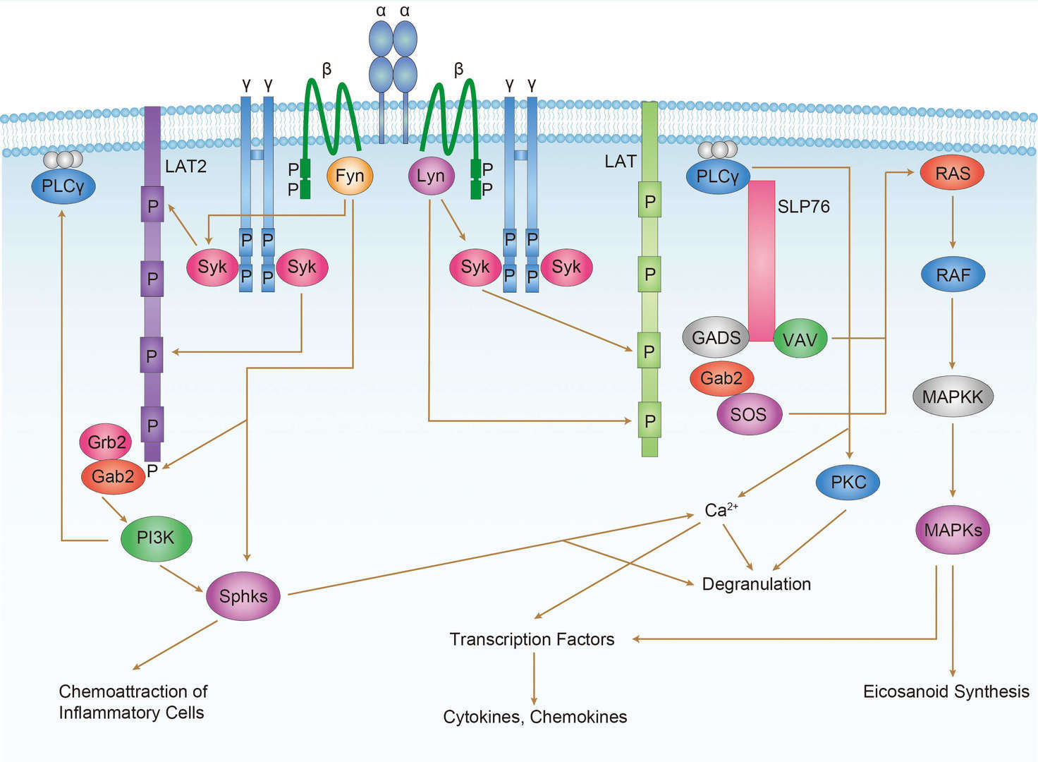
 FcεR1 Signaling Pathway
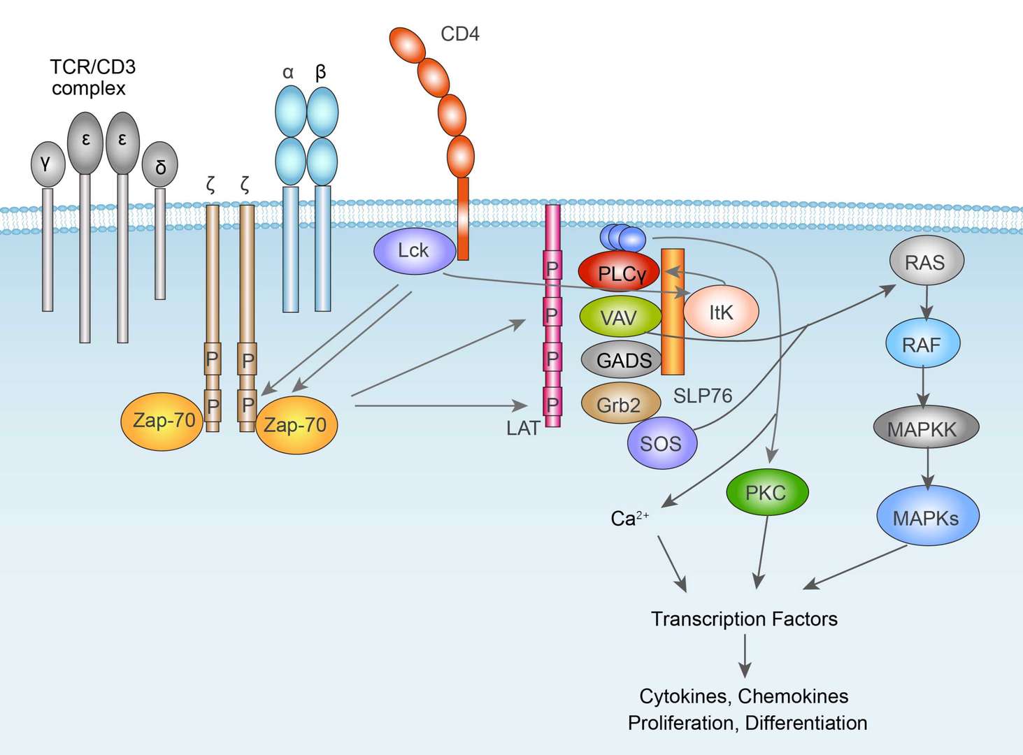
 TCR Signaling Pathway
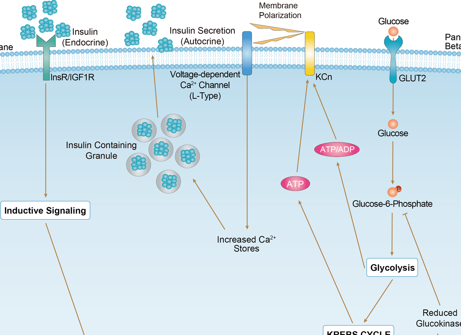
 Maturity Onset Diabetes of the Young


