

Anti-Human CD80 Recombinant Antibody (Galiximab)
CAT#: TAB-215
Recombinant monoclonal antibody to CD8. Galiximab is a monoclonal antibody designed for the treatment of B-cell lymphoma. As of September 2009, The drug is a chimeric antibody from Macaca irus and Homo sapiens.
Published Data
Gene Expression

Inhib
Figure 1 Galiximab inhibits cell proliferation of B-NHL cell lines and sensitizes B-NHL cells to apoptosis by CDDP and TRAIL.
A, surface expression of CD80 on B-NHL cell lines. The surface expression of CD80 was analyzed by flow cytometry. A, representative histogram is shown for the various B-NHL cell lines and an isotype control is shown. In addition, the mean fluorescence intensity (MFI) is represented in the table. B, sensitization to apoptosis. B-NHL cell lines were treated with galiximab (20 mg/mL) for 18 hours and followed by treatment with CDDP (5 mg/mL) or TRAIL (5 ng/mL) for an additional 18 hours and apoptosis was determined by activation of caspase-3 (left) as described. Ã, P < 0.05; ÃÃ, P < 0.01. Apoptosis was also determined by Annexin-V as described (right). C, galiximab-induced inhibition of Raji cell viability and cell recovery. The B-NHL cell line Raji was treated with various concentrations of galiximab and incubated for different time periods (6-24 hours), and cell viability was determined by trypan blue dye exclusion and total cell recovery was recorded. Raji cells that were not treated with galiximab represented 100% viability. The data represent the mean AE SD from 3 independent experiments Ã, P < 0.05. D, galiximab sensitizes resistant B-NHL cell lines to apoptosis by CDDP and TRAIL. Sensitization of B-NHL Raji cells by galiximab to apoptosis by CDDP or TRAIL is synergistic. Raji cells were treated with various concentrations of galiximab (10-100 mg/mL) for 18 hours and then treated with either CDDP (5, 10, and 20 mg/mL; left) or only one concentration of galiximab (20 mg/mL) and various concentrations of TRAIL (2.5, 5, and 10 ng/mL; right) for an additional 18 hours and apoptosis was determined. The data represent the mean AE SD from 3 independent experiments. Ã, P < 0.05; ÃÃ, P < 0.01. In addition, the data were analyzed for synergy by isobologram analysis as described in Materials and Methods. The isobologram is represented left of D. AAD, aminoactinomycin D.
Martinez-Paniagua, M. A., Vega, M. I., Huerta-Yepez, S., Baritaki, S., Vega, G. G., Hariharan, K., & Bonavida, B. (2012). Galiximab Signals B-NHL Cells and Inhibits the Activities of NF-κB–Induced YY1-and Snail-Resistant Factors: Mechanism of Sensitization to Apoptosis by Chemoimmunotherapeutic Drugs. Molecular cancer therapeutics, 11(3), 572-581.

WB
Figure 2 Galiximab inhibits NF-kB activity in Raji cells and the role of NF-kB inhibition in the sensitization of Raji to apoptosis by CDDP and TRAIL.
Raji cells were treated with different concentrations of galiximab (25, 50, and 100 mg/mL) for 18 hours, and aliquots were used to prepare both nuclear and total cell lysates as described in Materials and Methods. A, Western blot analysis for NF-kB expression. Total cell lysates were tested for various gene products of the NF-kB pathway. b-Actin was used as a loading control. Densitometric analysis is also shown and intensity of the bands was normalized to b-actin bands. B, inhibition of NF-kB DNA-binding activity by galiximab. Raji cells were treated with galiximab (25 mg/mL). Nuclear lysates were tested for NF-kB DNAbinding activity by EMSA as described. The NF-kB inhibitor DHMEQ (10 mg/mL) was used as a positive control and cold probes as competitors. For the supershift assay, the nuclear proteins were incubated with anti-p65 antibody overnight at 4 C before the analysis by EMSA. C, galiximab-induced inhibition of NF-kB in the sensitization to apoptosis by CDDP and TRAIL. Raji cells were treated with galiximab (20 mg/mL) for 18 hours or with the NF-kB inhibitor DHMEQ (10 mg/mL) for 18 hours and the cells were subsequently treated with either CDDP (5 mg/mL) or TRAIL (5 mg/mL) for an additional 24 hours and apoptosis was determined. The data represent the mean AE SD from 3 independent experiments. Ã, P < 0.01. D, inhibition of the AKT pathway by galiximab. Total cell lysates were tested for various gene products of the AKT pathway. b-Actin was used as a loading control. Densitometric analysis is also shown.
Martinez-Paniagua, M. A., Vega, M. I., Huerta-Yepez, S., Baritaki, S., Vega, G. G., Hariharan, K., & Bonavida, B. (2012). Galiximab Signals B-NHL Cells and Inhibits the Activities of NF-κB–Induced YY1-and Snail-Resistant Factors: Mechanism of Sensitization to Apoptosis by Chemoimmunotherapeutic Drugs. Molecular cancer therapeutics, 11(3), 572-581.

WB
Figure 3 Galiximab inhibits the expression and the activity of the transcription factors YY1 and Snail in Raji cells.
Inhibition of YY1 and Snail by galiximab. Raji cells were treated with various concentrations of galiximab (25, 50, and 100 mg/mL) for 18 hours and total cell lysates were prepared for Western blot analysis. b-Actin was used as a loading control. The Western blots analyses were also analyzed by densitometry and is shown below the Western blot analysis figure.
Martinez-Paniagua, M. A., Vega, M. I., Huerta-Yepez, S., Baritaki, S., Vega, G. G., Hariharan, K., & Bonavida, B. (2012). Galiximab Signals B-NHL Cells and Inhibits the Activities of NF-κB–Induced YY1-and Snail-Resistant Factors: Mechanism of Sensitization to Apoptosis by Chemoimmunotherapeutic Drugs. Molecular cancer therapeutics, 11(3), 572-581.

FC
Figure 4 Galiximab inhibits cell proliferation of B-NHL cell lines and sensitizes B-NHL cells to apoptosis by CDDP and TRAIL.
A, surface expression of CD80 on B-NHL cell lines. The surface expression of CD80 was analyzed by flow cytometry. A, representative histogram is shown for the various B-NHL cell lines and an isotype control is shown. In addition, the mean fluorescence intensity (MFI) is represented in the table. B, sensitization to apoptosis. B-NHL cell lines were treated with galiximab (20 μg/mL) for 18 hours and followed by treatment with CDDP (5 μg/mL) or TRAIL (5 ng/mL) for an additional 18 hours and apoptosis was determined by activation of caspase-3 (left) as described. *, P < 0.05; **, P < 0.01. Apoptosis was also determined by Annexin-V as described (right). C, galiximab-induced inhibition of Raji cell viability and cell recovery. The B-NHL cell line Raji was treated with various concentrations of galiximab and incubated for different time periods (6–24 hours), and cell viability was determined by trypan blue dye exclusion and total cell recovery was recorded. Raji cells that were not treated with galiximab represented 100% viability. The data represent the mean ± SD from 3 independent experiments *, P < 0.05. D, galiximab sensitizes resistant B-NHL cell lines to apoptosis by CDDP and TRAIL. Sensitization of B-NHL Raji cells by galiximab to apoptosis by CDDP or TRAIL is synergistic. Raji cells were treated with various concentrations of galiximab (10–100 μg/mL) for 18 hours and then treated with either CDDP (5, 10, and 20 μg/mL; left) or only one concentration of galiximab (20 μg/mL) and various concentrations of TRAIL (2.5, 5, and 10 ng/mL; right) for an additional 18 hours and apoptosis was determined. The data represent the mean ± SD from 3 independent experiments. *, P < 0.05; **, P < 0.01. In addition, the data were analyzed for synergy by isobologram analysis as described in Materials and Methods. The isobologram is represented left of D. AAD, aminoactinomycin D.
Martinez-Paniagua, M. A., Vega, M. I., Huerta-Yepez, S., Baritaki, S., Vega, G. G., Hariharan, K., & Bonavida, B. (2012). Galiximab Signals B-NHL Cells and Inhibits the Activities of NF-κB–Induced YY1-and Snail-Resistant Factors: Mechanism of Sensitization to Apoptosis by Chemoimmunotherapeutic Drugs. Molecular cancer therapeutics, 11(3), 572-581.

Activ
Figure 5 Galiximab inhibits the expression and the activity of the transcription factors YY1 and Snail in Raji cells.
A, inhibition of YY1 and Snail by galiximab. Raji cells were treated with various concentrations of galiximab (25, 50, and 100 μg/mL) for 18 hours and total cell lysates were prepared for Western blot analysis. β-Actin was used as a loading control. The Western blots analyses were also analyzed by densitometry and is shown below the Western blot analysis figure. B, galiximab inhibits the DNA-binding activity of YY1 and Snail. Raji cells were treated with galiximab (25 μg/mL) for 18 hours and nuclear lysates were tested for DNA-binding activities for YY1 and Snail as described in Materials and Methods. The specificity of DNA-binding activity was determined by the use of a corresponding competitive cold probe and in the absence of nuclear extracts in the assay.
Martinez-Paniagua, M. A., Vega, M. I., Huerta-Yepez, S., Baritaki, S., Vega, G. G., Hariharan, K., & Bonavida, B. (2012). Galiximab Signals B-NHL Cells and Inhibits the Activities of NF-κB–Induced YY1-and Snail-Resistant Factors: Mechanism of Sensitization to Apoptosis by Chemoimmunotherapeutic Drugs. Molecular cancer therapeutics, 11(3), 572-581.
❮


 ❯
❯

Normal Tissue
Figure 1 Bone marrow
Hematopoietic cells
Staining: Medium
Intensity: Strong
Quantity: <25%
Location: Cytoplasmic/membranous
* Image credit: Image credit: Human Protein Atlas https://v21.proteinatlas.org/images/25368/56116_B_6_4.jpg

Normal Tissue
Figure 2 Lymph node
Germinal center cells
Staining: Medium
Intensity: Strong
Quantity: <25%
Location: Cytoplasmic/membranous
Non-germinal center cells
Staining: Medium
Intensity: Strong
Quantity: <25%
Location: Cytoplasmic/membranous
* Image credit: Image credit: Human Protein Atlas https://v21.proteinatlas.org/images/25368/56076_A_9_8.jpg

RNA Expression
Figure 3 RNA cell line category: Cell line enhanced (Daudi, HDLM-2, Karpas-707, U-266/70)
Cell lines ordered by descending RNA expression order.
* Image credit: Image credit: Human Protein Atlas https://v21.proteinatlas.org/ENSG00000121594-CD80
❮


 ❯
❯
Specifications
- Immunogen
- CD80-transfected CHO cell line.
- Host Species
- primate
- Derivation
- Chimeric (primate/human)
- Type
- IgG1 - lambda
- Specificity
- Tested positive against native human antigen.
- Species Reactivity
- Human
- Applications
- WB, ELISA, FC, IP, FuncS, IF, Neut, Inhib, Activ
- CAS
- 357613-77-5
- Generic Name
- Galiximab
- UNII
- S9OX9692ZB
- Related Disease
- Non-Hodgkin's lymphoma (NHL)
Product Property
- Purity
- >95.0%, determined by analysis by RP-HPLC & analysis by SDS-PAGE.
- Storage
- Store at 4°C for up to 3 months. For longer term storage aliquot into small volumes and store at -20°C.
Applications
- Application Notes
- The CD80 antibody has been reported in applications of WB, ELISA, FC, IP, FuncS, IF, Neut, Inhib, Activ.
Target
- Alternative Names
- Galiximab;357613-77-5;IDEC-114;16C10;CD80;CD80 molecule;CD28LG, CD28LG1, CD80 antigen (CD28 antigen ligand 1, B7 1 antigen) , CD80 molecule;T-lymphocyte activation antigen CD80;B lymphocyte activation antigen B7;B7 1;B7.1;activation B7-1 antigen;costimula
- Gene ID
- 941
- UniProt ID
- P33681
REVIEWS AND Q&AS
CITATIONS
RESOURCES
DOWNLOADS
RELATED PRODUCTS
Inquiry
Navs
Customer Review

There are currently no Customer reviews or questions for TAB-215. Click the button above to contact us or submit your feedback about this product.
Submit Your Publication
Published with our product? Submit your paper and receive a 10% discount on your next order! Share your research to earn exclusive rewards.
Related Diseases
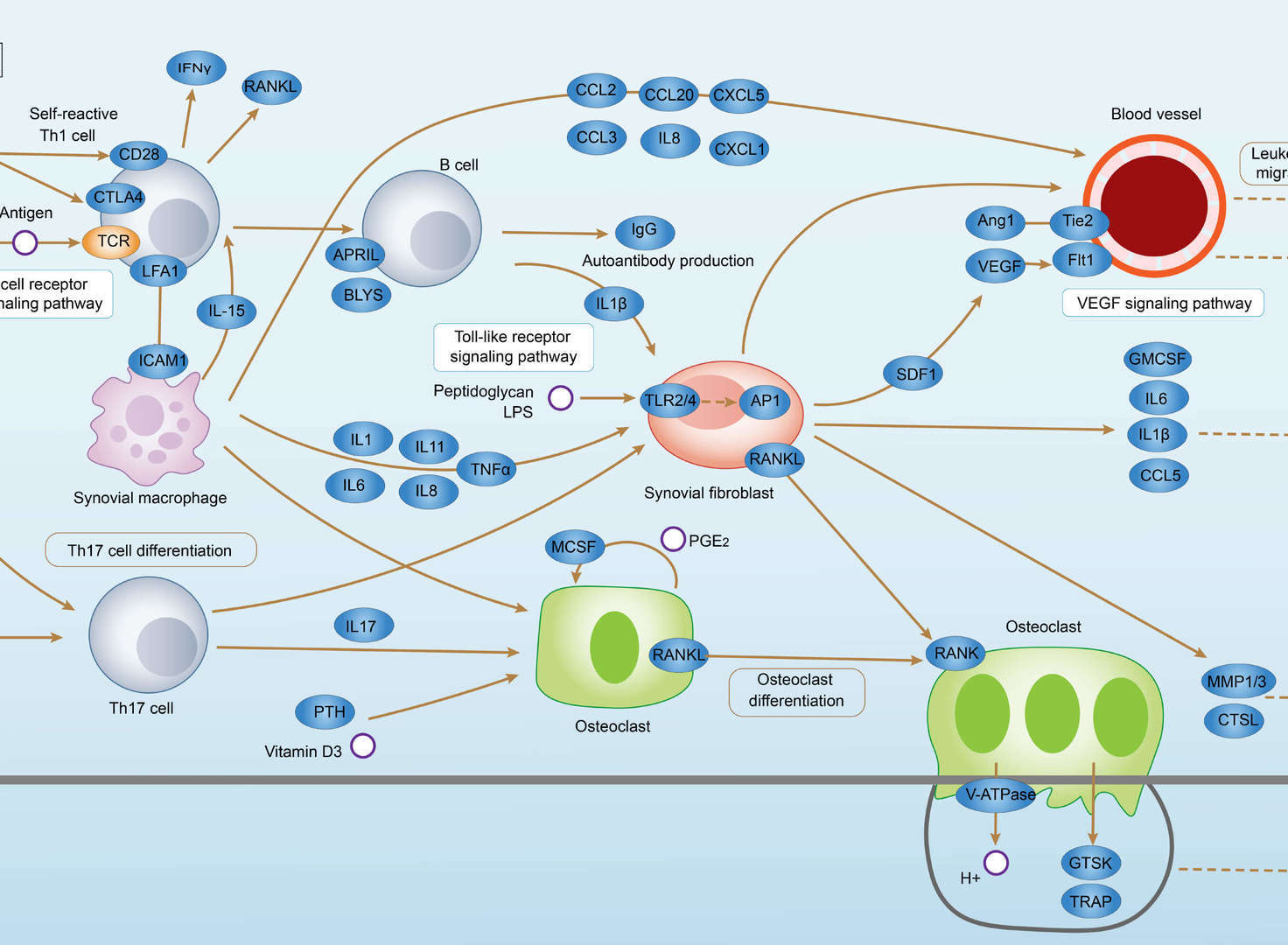 Rheumatoid Arthritis
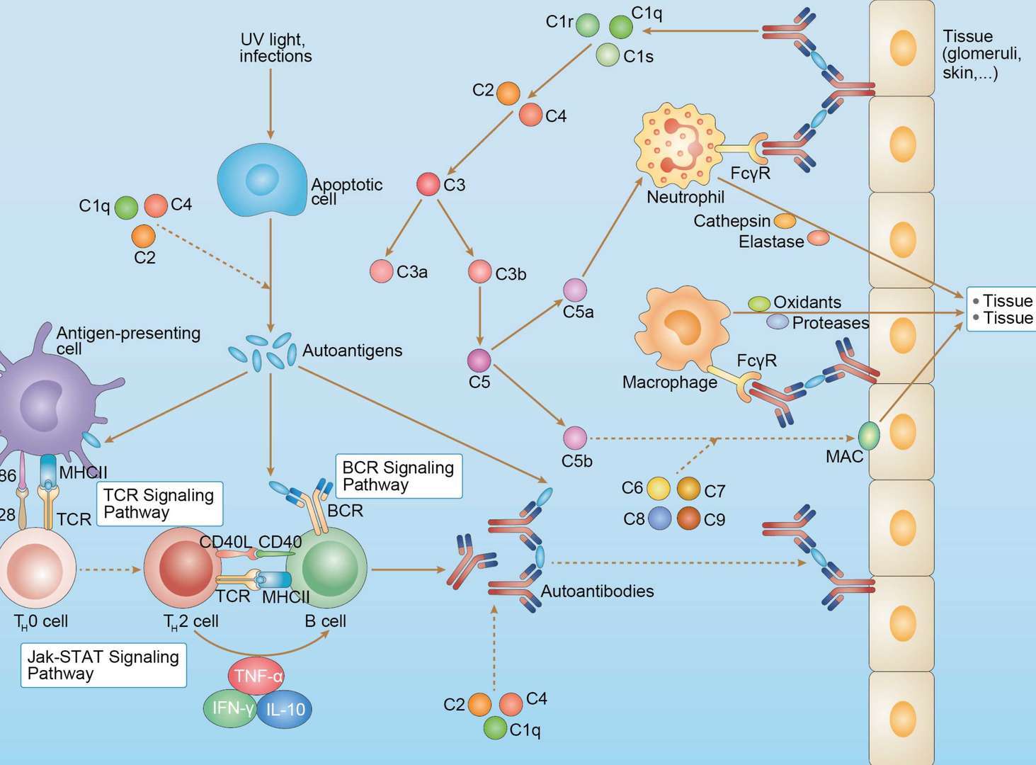 Systemic Lupus Erythematosus
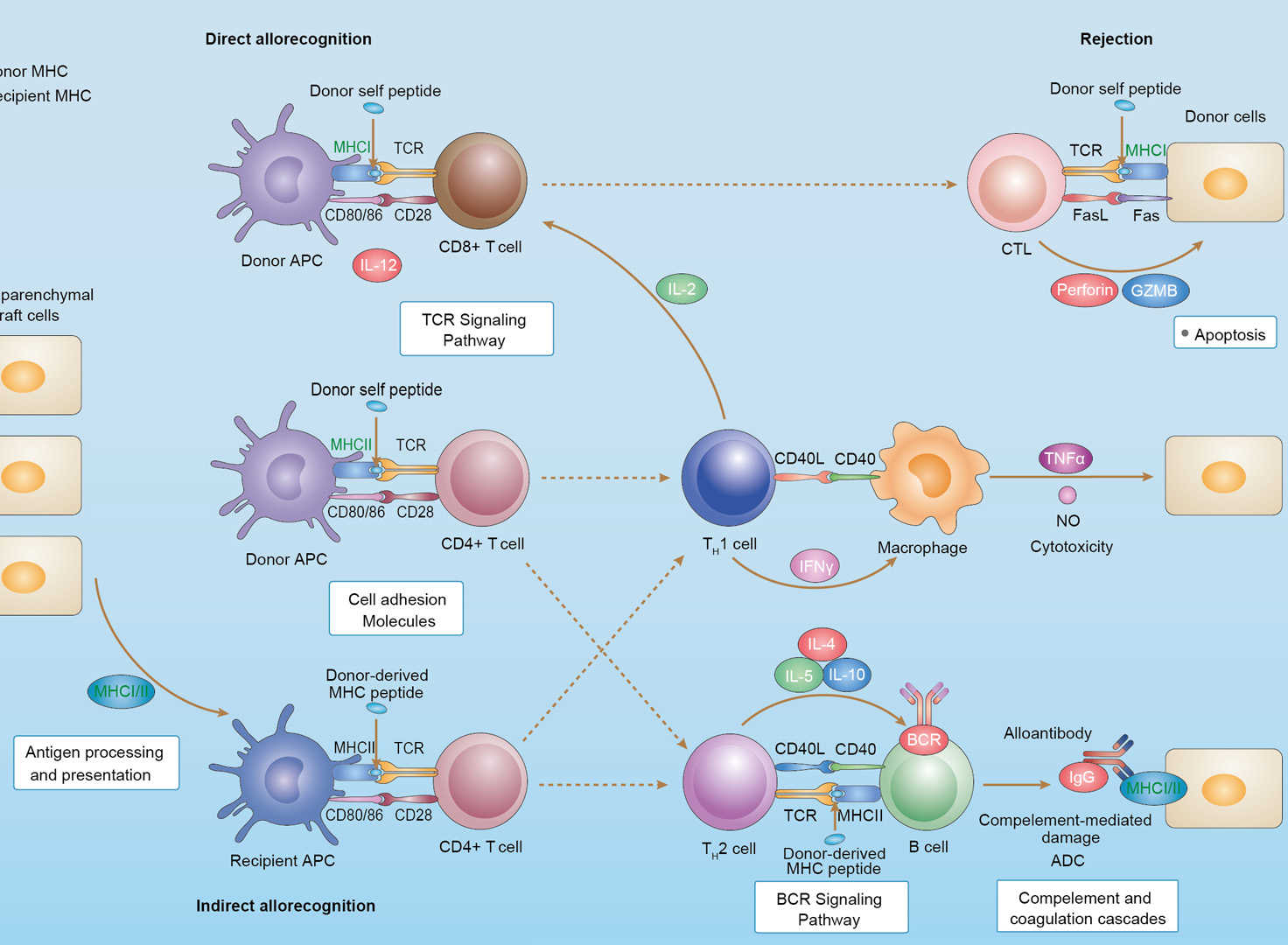 Allograft Rejection
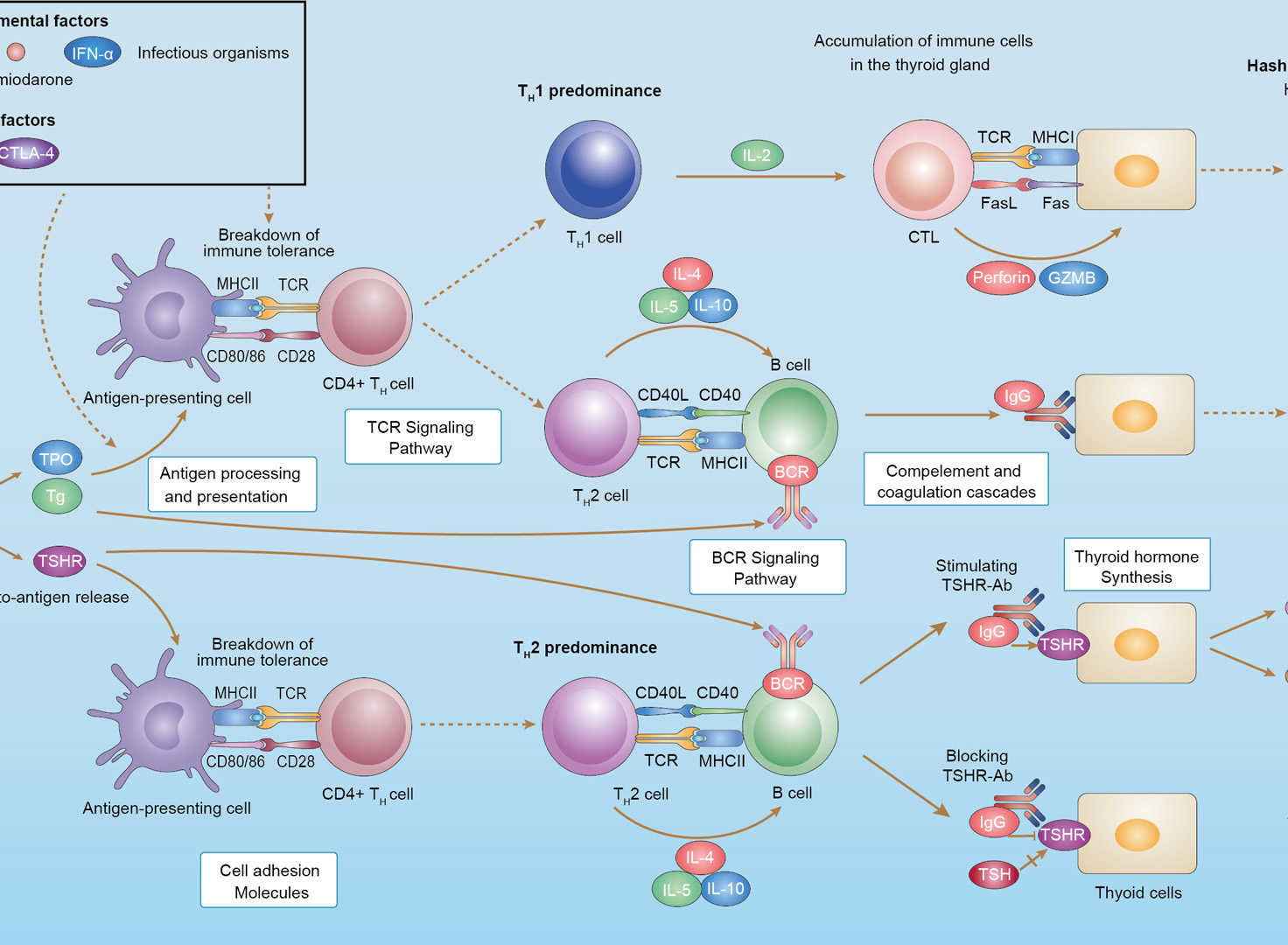 Autoimmune Thyroid Disease
 Graft-versus-Host Disease
 Maturity Onset Diabetes of the Young
Downloadable Resources
Download resources about recombinant antibody development and antibody engineering to boost your research.
Product Notes
This is a product of Creative Biolabs' Hi-Affi™ recombinant antibody portfolio, which has several benefits including:
• Increased sensitivity
• Confirmed specificity
• High repeatability
• Excellent batch-to-batch consistency
• Sustainable supply
• Animal-free production
See more details about Hi-Affi™ recombinant antibody benefits.
Datasheet
MSDS
COA
Certificate of Analysis LookupTo download a Certificate of Analysis, please enter a lot number in the search box below. Note: Certificate of Analysis not available for kit components.
Lot Number:
Protocol & Troubleshooting
We have outlined the assay protocols, covering reagents, solutions, procedures, and troubleshooting tips for common issues in order to better assist clients in conducting experiments with our products. View the full list of Protocol & Troubleshooting.
See other products for "Galiximab"
Afuco™ Anti-CD80 ADCC Recombinant Antibody, ADCC Enhanced (AFC-TAB-215)This product is an ADCC enhanced antibody produced by our Afuco™ platform. Recombinant monoclonal antibody to CD8. It is a monoclonal antibody designed for the treatment of B-cell lymphoma.
DrugMonitor™ Anti-Galiximab Antibody (VS-1224-YC502)Galiximab is a chimeric monoclonal antibody used as an immunosuppressive drug. The DrugMonitor™ Anti-Galiximab Antibody (VS-1224-YC502) is an anti-drug antibody (ADA) against Galiximab. This drug-based antibody is raised in mice immunized with the Galiximab. The anti-Galiximab antibody may be used in ELISA, pharmacokinetics (PK), and pharmacodynamics (PD) analyses, or serves as a reference standard in ADA assays. It also is an excellent tool for therapeutic drug monitoring, allowing to evaluate the drug efficacy and determine the drug concentration of the Galiximab in samples.
See other products for "CD80"
Select a product category from the dropdown menu below to view related products.
| CAT | Product Name | Application | Type |
|---|---|---|---|
| MOB-1234z | Mouse Anti-CD80 Recombinant Antibody (clone 31G9) | WB, FC, IHC | Mouse IgG2b, κ |
| CAT | Product Name | Application | Type |
|---|---|---|---|
| AGTO-L001G | anti-CD80 immunotoxin 6G7 (IgG)-Gel | Cytotoxicity assay, Functional assay |
| CAT | Product Name | Application | Type |
|---|---|---|---|
| AGTO-L001S | anti-CD80 immunotoxin 6G7 (IgG)-Sap | Cytotoxicity assay, Functional assay |
| CAT | Product Name | Application | Type |
|---|---|---|---|
| AGTO-L001O | anti-CD80 immunotoxin 6G7 (IgG)-Bouganin | Cytotoxicity assay, Functional assay |
| CAT | Product Name | Application | Type |
|---|---|---|---|
| TAB-347LC | Mouse Anti-CD80 Recombinant Antibody (TAB-347LC) | FC | Mouse IgG |
| CAT | Product Name | Application | Type |
|---|---|---|---|
| TAB-347LC-S(P) | Mouse Anti-CD80 Recombinant Antibody; scFv Fragment (TAB-347LC-S(P)) | FC | Mouse scFv |
| CAT | Product Name | Application | Type |
|---|---|---|---|
| TAB-347LC-F(E) | Mouse Anti-CD80 Recombinant Antibody; Fab Fragment (TAB-347LC-F(E)) | FC | Mouse Fab |
| CAT | Product Name | Application | Type |
|---|---|---|---|
| Gly-159LC | Recombinant Anti-Human CD80 Antibody (Fc glycosylation/Non fucosylated) | ELISA | Chimeric antibody (primate/human) |
| Gly-159LC-1 | Recombinant Anti-Human CD80 Antibody (Fc glycosylation/Non fucosylated) | ELISA | Chimeric antibody (primate/human) |
| CAT | Product Name | Application | Type |
|---|---|---|---|
| MOB-0490CT | Recombinant Mouse anti-Human CD80 Monoclonal antibody (NFN-344) | FC |
| CAT | Product Name | Application | Type |
|---|---|---|---|
| BRD-0066MZ | Chicken Anti-BB1 Polyclonal IgY | WB | Chicken antibody |
| CAT | Product Name | Application | Type |
|---|---|---|---|
| NEUT-409CQ | Mouse Anti-CD80 Recombinant Antibody (clone CBL764) | FC, IHC, CyTOF®, ELISA, Neut | Mouse IgG1 |
| CAT | Product Name | Application | Type |
|---|---|---|---|
| NEUT-410CQ | Mouse Anti-CD80 Recombinant Antibody (clone 6N36) | FC, IHC, Neut, WB | Mouse IgG1 |
| CAT | Product Name | Application | Type |
|---|---|---|---|
| NEUT-411CQ | Mouse Anti-CD80 Recombinant Antibody (clone AS29) | IP, Neut | Mouse IgG1 |
| CAT | Product Name | Application | Type |
|---|---|---|---|
| NEUT-412CQ | Mouse Anti-CD80 Recombinant Antibody (VMC172) | Neut, FC | Mouse IgG1 |
| CAT | Product Name | Application | Type |
|---|---|---|---|
| NEUT-413CQ | Rat Anti-Cd80 Recombinant Antibody (clone 1G10) | IP, Block | Rat IgG2a, κ |
| CAT | Product Name | Application | Type |
|---|---|---|---|
| NEUT-414CQ | Hamster Anti-Cd80 Recombinant Antibody (NEUT-414CQ) | Block, FC, IHC-Fr | Hamster IgG |
| CAT | Product Name | Application | Type |
|---|---|---|---|
| NEUT-415CQ | Hamster Anti-Cd80 Recombinant Antibody (clone 16-10A1) | FC, FuncS, Neut, IHC, IHC-Fr | Hamster IgG |
| CAT | Product Name | Application | Type |
|---|---|---|---|
| NEUT-416CQ | Mouse Anti-Cd80 Recombinant Antibody (clone 3H5) | Block, FC | Mouse IgG1 |
| CAT | Product Name | Application | Type |
|---|---|---|---|
| MOR-0583 | Hi-Affi™ Rabbit Anti-CD80 Recombinant Antibody (clone DS583AB) | WB, IHC | Rabbit IgG |
| CAT | Product Name | Application | Type |
|---|---|---|---|
| MRO-0318-CN | Rabbit Anti-CD80 Recombinant Antibody (clone JF100-4) | WB, IHC | Rabbit IgG |
| CAT | Product Name | Application | Type |
|---|---|---|---|
| MRO-0319-CN | Mouse Anti-CD80 Recombinant Antibody (clone 8-E5) | WB, IF, IHC, FC | Mouse IgG2b |
| CAT | Product Name | Application | Type |
|---|---|---|---|
| MRO-1762-CN | Rabbit Anti-CD80 Polyclonal Antibody (MRO-1762-CN) | WB, IF, IHC, FC | Rabbit IgG |
| CAT | Product Name | Application | Type |
|---|---|---|---|
| AFC-TAB-215 | Afuco™ Anti-CD80 ADCC Recombinant Antibody, ADCC Enhanced (AFC-TAB-215) | ELISA, FC, IP, FuncS, IF | ADCC enhanced antibody |
| CAT | Product Name | Application | Type |
|---|---|---|---|
| FN-040CQ | Mouse Anti-CD80 Recombinant Antibody (FN-040CQ) | FC, ELISA, WB, Block | Mouse IgM, κ |
| CAT | Product Name | Application | Type |
|---|---|---|---|
| HPAB-0063-YJ-S(P) | Human Anti-CD80 Recombinant Antibody (clone hu1F1); scFv Fragment | ELISA, FuncS | Humanized scFv |
| CAT | Product Name | Application | Type |
|---|---|---|---|
| HPAB-0063-YJ-F(E) | Human Anti-CD80 Recombinant Antibody (clone hu1F1); Fab Fragment | ELISA, FuncS | Humanized Fab |
| CAT | Product Name | Application | Type |
|---|---|---|---|
| HPAB-0708-FY-F(E) | Mouse Anti-CD80 Recombinant Antibody (clone AB007); scFv Fragment | ELISA | Mouse scFv |
| CAT | Product Name | Application | Type |
|---|---|---|---|
| HPAB-0708-FY-S(P) | Mouse Anti-CD80 Recombinant Antibody (clone AB007); Fab Fragment | ELISA | Mouse Fab |
| CAT | Product Name | Application | Type |
|---|---|---|---|
| HPAB-1587WJ-F(E) | Mouse Anti-CD80 Recombinant Antibody; Fab Fragment (clone 1F1) | ELISA, WB | Mouse Fab |
| CAT | Product Name | Application | Type |
|---|---|---|---|
| HPAB-1587WJ-S(P) | Mouse Anti-CD80 Recombinant Antibody; scFv Fragment (clone 1F1) | ELISA, WB | Mouse scFv |
| CAT | Product Name | Application | Type |
|---|---|---|---|
| VS-0424-XY56 | AbPlus™ Anti-CD80 Magnetic Beads (AS29) | IP, Protein Purification |
| CAT | Product Name | Application | Type |
|---|---|---|---|
| VS-0225-XY73 | CytoStream™ Mouse Anti-CD80 Recombinant Antibody (clone 2D10.4) | FC | Mouse IgG1, kappa |
| CAT | Product Name | Application | Type |
|---|---|---|---|
| VS13-YC175 | CytoStream™ Hamster Anti-Mouse Cd80 Recombinant Antibody (VS13-YC175) | FC | Armenian hamster IgG |
| CAT | Product Name | Application | Type |
|---|---|---|---|
| VS13-YC176 | CytoStream™ Rabbit Anti-CD80 Recombinant Antibody (VS13-YC176) | FC, ELISA | Rabbit IgG |
| CAT | Product Name | Application | Type |
|---|---|---|---|
| VS13-YC177 | CytoStream™ Mouse Anti-CD80 Recombinant Antibody (VS13-YC177) | WB, IHC-P, FC, ICC | Mouse IgG |
| CAT | Product Name | Application | Type |
|---|---|---|---|
| VS-0425-FY9 | Mouse Anti-CD80 (clone AB007) scFv-Fc Chimera | IA | Mouse IgG1, scFv-Fc |
| CAT | Product Name | Application | Type |
|---|---|---|---|
| VS-0425-YC176 | Recombinant Anti-CD80 Vesicular Antibody, EV Displayed (VS-0425-YC176) | ELISA, FC, Cell-uptake |
| CAT | Product Name | Application | Type |
|---|---|---|---|
| VS-0525-XY1224 | Anti-Mouse CD80 Immunohistochemistry Kit | IHC |
| CAT | Product Name | Application | Type |
|---|---|---|---|
| VS-0825-YC70 | SmartAb™ Recombinant Anti-CD80 pH-dependent Antibody (Clone Galiximab) | WB, ELISA, FC, IP IF, Neut | IgG1 lambda (combining the Macaque antibody's antigen-binding variable regions with a human IgG1 constant region) |
Specific Inquiry

See Our Custom Production in Action


Popular Products

Application: ELISA, IP, FC, FuncS, Neut, IF, ICC

Application: ELISA, FC, IP, FuncS, IF, Neut, ICC

Application: WB, IF, IP, Neut, FuncS, ELISA, FC

Application: Neut, ELISA, IF, IP, FuncS, FC, IHC

Application: FuncS, IF, Neut, ELISA, FC, IP, IHC

Application: IF, IP, Neut, FuncS, ELISA, FC, WB

Application: FC, IP, ELISA, Neut, FuncS, IF, ICC

Application: Neut, ELISA, IF, IP, FuncS, FC, ICC

Application: FC, IP, ELISA, Neut, FuncS, IF, WB

Application: Inhib, Cyt

Application: FuncS, IF, Neut, ELISA, FC, IP, ICC

Application: IF, IP, Neut, FuncS, ELISA, FC, ICC
-2-1.png)
Application: IP, IF, FuncS, FC, Neut, ELISA, IHC

Application: WB, Neut, ELISA, IF, IP, FuncS, FC


Application: Block, Cyt, FuncS, Inhib


For research use only. Not intended for any clinical use. No products from Creative Biolabs may be resold, modified for resale or used to manufacture commercial products without prior written approval from Creative Biolabs.
Send Inquiry
This site is protected by reCAPTCHA and the Google Privacy Policy and Terms of Service apply.