Rabbit Anti-ERBB3 Recombinant Antibody (clone R04-8J6)
CAT#: VS3-XY3348
It is a recombinant rabbit antibody targeting the human ERBB3 (erb-b2 receptor tyrosine kinase 3), which involves in the regulation of myeloid cell differentiation. This antibody reacts with human and rat ERBB3. It has been determined using Western Blot and Immunoprecipitation. The recommended dilutions of Western Blot and Immunoprecipitation is 1/500-1/1000 and 1/20, respectively.
Gene Expression

Subcellular Location and Protein Expression
Figure 1 IF staining of human cell line U-2 OS
Immunofluorescent staining of human cell line U-2 OS shows localization to plasma membrane & actin filaments.
* Image credit: Image credit: Human Protein Atlas https://v21.proteinatlas.org/images/25331/if_selected.jpg

Subcellular Location and Protein Expression
Figure 2 IHC staining of human testis
Immunohistochemical staining of human testis shows strong cytoplasmic positivity in cells in seminiferus ducts and Leydig cells.
* Image credit: Image credit: Human Protein Atlas https://v21.proteinatlas.org/images/45396/ihc_selected.jpg

Subcellular Location and Protein Expression
Figure 3 IHC staining of human small intestine
Immunohistochemical staining of human small intestine shows strong cytoplasmic positivity in glandular cells.
* Image credit: Image credit: Human Protein Atlas https://v21.proteinatlas.org/images/70524/162441_A_4_2_selected.jpg

Subcellular Location and Protein Expression
Figure 4 IHC staining of human kidney
Immunohistochemical staining of human kidney shows strong cytoplasmic and membranous positivity in renal tubular cells.
* Image credit: Image credit: Human Protein Atlas https://v21.proteinatlas.org/images/25331/67596_A_8_5_selected.jpg

Subcellular Location and Protein Expression
Figure 5 IF staining of human cell line A-431
Immunofluorescent staining of human cell line A-431 shows localization to plasma membrane.
* Image credit: Image credit: Human Protein Atlas https://v21.proteinatlas.org/images/25331/659_B11_4_blue_red_green.jpg

Subcellular Location and Protein Expression
Figure 6 IF staining of human cell line U-251 MG
Immunofluorescent staining of human cell line U-251 MG shows localization to plasma membrane.
* Image credit: Image credit: Human Protein Atlas https://v21.proteinatlas.org/images/25331/658_B11_1_blue_red_green.jpg

Normal Tissue
Figure 7 Cerebral cortex
Endothelial cells
Staining:Medium
Intensity: Moderate
Quantity:>75%
Location: Cytoplasmic/membranous
Neuronal cells
Staining:High
Intensity: Strong
Quantity:>75%
Location: Cytoplasmic/membranous nuclear
Neuropil
Staining:Medium
Intensity: Moderate
Quantity:>75%
Location: Cytoplasmic/membranous
* Image credit: Image credit: Human Protein Atlas https://v21.proteinatlas.org/images/45396/115200_B_7_5.jpg

Normal Tissue
Figure 8 Colon
Endothelial cells
Staining:Medium
Intensity: Moderate
Quantity:>75%
Location: Cytoplasmic/membranous
Glandular cells
Staining:Medium
Intensity: Strong
Quantity: <25%
Location: Cytoplasmic/membranous
Peripheral nerve/ganglion
Staining:Medium
Intensity: Moderate
Quantity:>75%
Location: Cytoplasmic/membranous
* Image credit: Image credit: Human Protein Atlas https://v21.proteinatlas.org/images/25331/67596_A_9_3.jpg

Normal Tissue
Figure 9 Liver
Cholangiocytes
Staining:Medium
Intensity: Moderate
Quantity:>75%
Location: Cytoplasmic/membranous
Hepatocytes
Staining:Medium
Intensity: Moderate
Quantity:>75%
Location: Cytoplasmic/membranous
* Image credit: Image credit: Human Protein Atlas https://v21.proteinatlas.org/images/45396/114856_A_8_4.jpg

Normal Tissue
Figure 10 Kidney
Cells in glomeruli
Staining:Medium
Intensity: Moderate
Quantity:>75%
Location: Cytoplasmic/membranous
Cells in tubules
Staining:High
Intensity: Strong
Quantity:>75%
Location: Cytoplasmic/membranous
* Image credit: Image credit: Human Protein Atlas https://v21.proteinatlas.org/images/45396/114856_A_9_5.jpg

Normal Tissue
Figure 11 Testis
Cells in seminiferous ducts
Staining:High
Intensity: Strong
Quantity:>75%
Location: Cytoplasmic/membranous
Leydig cells
Staining:High
Intensity: Strong
Quantity:>75%
Location: Cytoplasmic/membranous
* Image credit: Image credit: Human Protein Atlas https://v21.proteinatlas.org/images/45396/114856_A_4_6.jpg

Normal Tissue
Figure 12 Skin
Fibroblasts
Staining:High
Intensity: Strong
Quantity:>75%
Location: Cytoplasmic/membranous
Keratinocytes
Staining:High
Intensity: Strong
Quantity:>75%
Location: Cytoplasmic/membranous
Langerhans
Staining:Medium
Intensity: Moderate
Quantity:>75%
Location: Cytoplasmic/membranous
Melanocytes
Staining:Medium
Intensity: Moderate
Quantity:>75%
Location: Cytoplasmic/membranous
* Image credit: Image credit: Human Protein Atlas https://v21.proteinatlas.org/images/45396/115200_B_8_1.jpg

Normal Tissue
Figure 13 Lymph node
Non-germinal center cells
Staining:High
Intensity: Strong
Quantity: 75%-25%
Location: Cytoplasmic/membranous
* Image credit: Image credit: Human Protein Atlas https://v21.proteinatlas.org/images/45396/114856_A_9_8.jpg

RNA Expression
Figure 14 RNA cell line category: Cell line enhanced (Hep G2, OE19, RT4, SK-BR-3, SK-MEL-30, T-47d, WM-115)
Cell lines ordered by descending RNA expression order.
* Image credit: Image credit: Human Protein Atlas https://v21.proteinatlas.org/ENSG00000065361-ERBB3
❮


 ❯
❯
Specifications
- Host Species
- Rabbit
- Type
- Rabbit IgG
- Specificity
- Human and Rat ERBB3
- Species Reactivity
- Human, Rat
- Clone
- R04-8J6
- Applications
- WB, IP
- Related Disease
- Lethal Congenital Contracture Syndrome 2
Product Property
- Clonality
- Monoclonal
- Aggregation
- < 5%
- Format
- Liquid
- Buffer
- PBS (pH 7.4), 150 mM NaCl and 50% glycerol
- Preservative
- 0.02% sodium azide
- Storage
- Store at 4°C short term. Aliquot and store at -20°C long term. Avoid freeze/thaw cycles.
- Shipping
- Ice packs
Applications
- Application Notes
- WB: 1/500-1/1000
IP: 1/20
Target
- Alternative Names
- HER3; FERLK; LCCS2; VSCN1; ErbB-3; c-erbB3; erbB3-S; MDA-BF-1; c-erbB-3; p180-ErbB3; p45-sErbB3; p85-sErbB3
- Gene ID
- 2065
- UniProt ID
- P21860
- Long Name
- erb-b2 receptor tyrosine kinase 3
- Cellular Localization
- Cell membrane
- Post Translation Modifications
- Glycosylation at Asn126, Asn250, Asn353, Asn408, Asn414, Asn437, Asn469, Asn522, Asn566, and Asn616 (NX_P21860 [NX_P21860-1])
Modification sites at PhosphoSitePlus (P21860)
Glycosylation from GlyConnect ERBB3_HUMAN (1711)
Glycosylation from GlyGen (P21860) 10 sites, 5 N-linked glycans (3 sites)
- Protein Refseq
- XP_047284456.1; XP_047284457.1
- Function
- Tyrosine-protein kinase that plays an essential role as cell surface receptor for neuregulins.
Binds to neuregulin-1 (NRG1) and is activated by it; ligand-binding increases phosphorylation on tyrosine residues and promotes its association with the p85 subunit of phosphatidylinositol 3-kinase (PubMed:20682778).
May also be activated by CSPG5 (PubMed:15358134).
Involved in the regulation of myeloid cell differentiation.
REVIEWS AND Q&AS
CITATIONS
RESOURCES
DOWNLOADS
RELATED PRODUCTS
Inquiry
Navs
Customer Review

There are currently no Customer reviews or questions for VS3-XY3348. Click the button above to contact us or submit your feedback about this product.
Submit Your Publication
Published with our product? Submit your paper and receive a 10% discount on your next order! Share your research to earn exclusive rewards.
Related Diseases
 EGFR Tyrosine Kinase Inhibitor Resistance
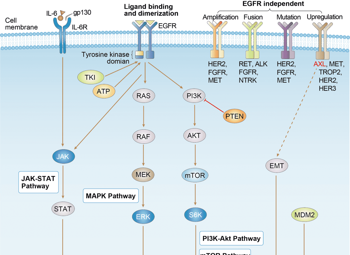
Related Signaling Pathways
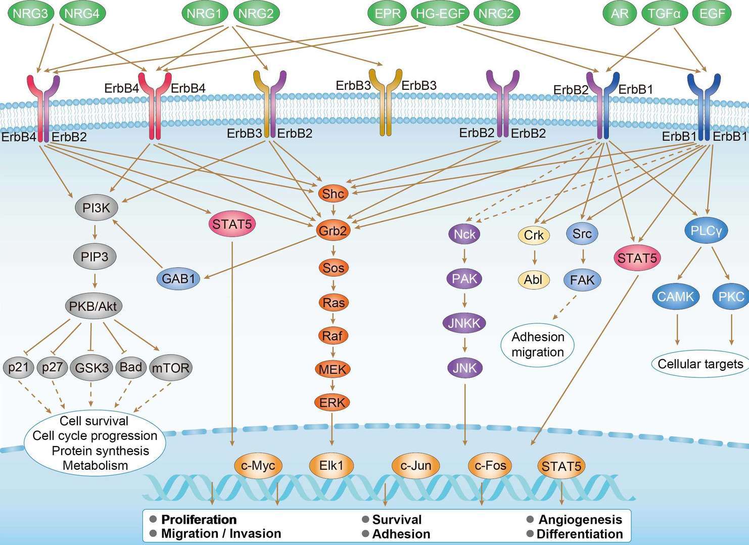 ErbB Signaling Pathway
Downloadable Resources
Download resources about recombinant antibody development and antibody engineering to boost your research.
Product Notes
This is a product of Creative Biolabs' Hi-Affi™ recombinant antibody portfolio, which has several benefits including:
• Increased sensitivity
• Confirmed specificity
• High repeatability
• Excellent batch-to-batch consistency
• Sustainable supply
• Animal-free production
See more details about Hi-Affi™ recombinant antibody benefits.
Datasheet
MSDS
COA
Certificate of Analysis LookupTo download a Certificate of Analysis, please enter a lot number in the search box below. Note: Certificate of Analysis not available for kit components.
Lot Number:
Isotype Control
- CAT
- Product Name
Recommended Dilution Buffer
- CAT
- Product Name
Secondary Antibody
- CAT
- Product Name
See other products for "ERBB3"
Select a product category from the dropdown menu below to view related products.
| CAT | Product Name | Application | Type |
|---|---|---|---|
| NAB-1729-sdAb | Recombinant Anti-human ERBB3 VHH Single Domain Antibody | WB, ICC, ChiP, FA, ELISA | Llama VHH |
| TAB-070CT | Llama Anti-ERBB3 Recombinant Single Domain Antibody (TAB-070CT) | FC, Block | Llama VHH |
| PABC-556 | Recombinant Llama Anti-ERBB3 Single Domain Antibody (PABC-556) | ELISA, SPR | Llama VHH |
| CAT | Product Name | Application | Type |
|---|---|---|---|
| MOB-1319z | Mouse Anti-ERBB3 Recombinant Antibody (clone 22C5) | ELISA, ICC, IF, WB | Mouse IgG1 |
| PABL-086 | Human Anti-ERBB3 Recombinant Antibody (clone KTN3379) | ELISA, WB, IF, FuncS | Human IgG |
| PABL-127 | Human Anti-ERBB3 Recombinant Antibody (PABL-127) | ELISA, WB, FuncS | Human IgG |
| PABL-128 | Mouse Anti-ERBB3 Recombinant Antibody (PABL-128) | WB, Block, FuncS | Mouse IgG |
| HPAB-0286-YC | Mouse Anti-ERBB3 Recombinant Antibody (clone 1153) | ELISA, FuncS, FC | Mouse IgG |
| CAT | Product Name | Application | Type |
|---|---|---|---|
| AGTO-L074E | HRGβ2-PE immunotoxin | Cytotoxicity assay, Functional assay |
| CAT | Product Name | Application | Type |
|---|---|---|---|
| PSBL-086 | Human Anti-ERBB3 Recombinant Antibody (clone KTN3379); scFv Fragment | ELISA, WB, IF, FuncS | Human scFv |
| PSBL-127 | Human Anti-ERBB3 Recombinant Antibody; scFv Fragment (PSBL-127) | ELISA, WB, FuncS | Human scFv |
| PSBL-128 | Mouse Anti-ERBB3 Recombinant Antibody; scFv Fragment (PSBL-128) | WB, Block, FuncS | Mouse scFv |
| HPAB-0286-YC-S(P) | Mouse Anti-ERBB3 Recombinant Antibody (clone 1153); scFv Fragment | ELISA, FuncS, FC | Mouse scFv |
| HPAB-0287-YC-S(P) | Mouse Anti-ERBB3 Recombinant Antibody (clone 920104); scFv Fragment | ELISA, FuncS, FC | Mouse scFv |
| CAT | Product Name | Application | Type |
|---|---|---|---|
| TAB-0033CL | Human Anti-ERBB3 Recombinant Antibody (TAB-0033CL) | ELISA, FuncS, Inhib, FC | Human IgG |
| TAB-0034CL | Human Anti-ERBB3 Recombinant Antibody (TAB-0034CL) | ELISA, FuncS, Inhib, FC | Human IgG |
| TAB-0035CL | Human Anti-ERBB3 Recombinant Antibody (TAB-0035CL) | ELISA, FuncS, Inhib, FC | Human IgG |
| TAB-0036CL | Human Anti-ERBB3 Recombinant Antibody (TAB-0036CL) | ELISA, FuncS, Inhib, FC | Human IgG |
| TAB-0037CL | Human Anti-ERBB3 Recombinant Antibody (TAB-0037CL) | ELISA, FuncS, Inhib, FC | Human IgG |
| CAT | Product Name | Application | Type |
|---|---|---|---|
| TAB-0552CL-S(P) | Mouse Anti-ERBB3 Recombinant Antibody; scFv Fragment (TAB-0552CL-S(P)) | ELISA, Inhib | Mouse scFv |
| TAB-0553CL-S(P) | Mouse Anti-ERBB3 Recombinant Antibody; scFv Fragment (TAB-0553CL-S(P)) | ELISA, Inhib | Mouse scFv |
| TAB-060CT-F(E) | Rabbit Anti-ERBB3 Recombinant Antibody; Fab Fragment (TAB-060CT-F(E)) | Inhib, ELISA | Rabbit Fab |
| TAB-064CT-F(E) | Mouse Anti-ERBB3 Recombinant Antibody; Fab Fragment (TAB-064CT-F(E)) | ELISA, FC | Mouse Fab |
| TAB-067CT-F(E) | Anti-Human HER3 Recombinant Antibody Fab Fragment (5101) | ELISA, Inhibition, FC |
| CAT | Product Name | Application | Type |
|---|---|---|---|
| TAB-057CT | Human Anti-ERBB3 Recombinant Antibody (TAB-057CT) | Inhibion, ELISA | Humanized antibody |
| TAB-057CT-S(P) | Human Anti-ERBB3 Recombinant Antibody; scFv Fragment (TAB-057CT-S(P)) | Inhibion, ELISA | Humanized scFv |
| TAB-058CT-S(P) | Human Anti-ERBB3 Recombinant Antibody; scFv Fragment (TAB-058CT-S(P)) | WB, ELISA, Inhib | Human scFv |
| TAB-059CT-S(P) | Anti-Human HER3 Recombinant Antibody scFv Fragment (LMAb3) | WB, ELISA, Inhibition, FC | Humanized antibody |
| TAB-063CT-S(P) | Human Anti-ERBB3 Recombinant Antibody; scFv Fragment (TAB-063CT-S(P)) | ELISA, Inhib, FC | Human scFv |
| CAT | Product Name | Application | Type |
|---|---|---|---|
| TAB-061CT | Human Anti-ERBB3 Recombinant Antibody (TAB-061CT) | Inhib, ELISA | Chimeric (Rabbit/Human) antibody |
| TAB-061CT-S(P) | Human Anti-ERBB3 Recombinant Antibody; scFv Fragment (TAB-061CT-S(P)) | Inhib, ELISA | Human scFv |
| TAB-061CT-F(E) | Human Anti-ERBB3 Recombinant Antibody; Fab Fragment (TAB-061CT-F(E)) | Inhib, ELISA | Chimeric (Rabbit/Human) Fab |
| CAT | Product Name | Application | Type |
|---|---|---|---|
| NEUT-738CQ | Mouse Anti-ERBB3 Recombinant Antibody (clone CBL932) | Neut, IP | Mouse IgG1 |
| NEUT-739CQ | Mouse Anti-ERBB3 Recombinant Antibody (clone CBL483) | FC, CyTOF®, ELISA, Neut | Mouse IgG1 |
| NEUT-740CQ | Human Anti-ERBB3 Recombinant Antibody (clone CBL1019) | Neut | Human IgG1, κ |
| NEUT-741CQ | Human Anti-ERBB3 Recombinant Antibody (clone CBL1020) | Neut | Human IgG1, κ |
| NEUT-742CQ | Mouse Anti-ERBB3 Recombinant Antibody (NEUT-742CQ) | Neut | Mouse IgG1 |
| CAT | Product Name | Application | Type |
|---|---|---|---|
| MOR-1179 | Hi-Affi™ Rabbit Anti-ERBB3 Recombinant Antibody (clone DS1179AB) | ICC, IF, WB | Rabbit IgG |
| CAT | Product Name | Application | Type |
|---|---|---|---|
| HPAB-0220-FY-F(E) | Human Anti-ERBB3 Recombinant Antibody (clone Hu-E14); Fab Fragment | ELISA, WB | Humanized Fab |
| HPAB-0171-WJ-F(E) | Human Anti-ERBB3 Recombinant Antibody; Fab Fragment (HPAB-0171-WJ-F(E)) | ELISA | Human Fab |
| HPAB-0200-WJ-F(E) | Mouse Anti-ERBB3 Recombinant Antibody (clone 205.10.1); Fab Fragment | ELISA, WB | Mouse Fab |
| HPAB-AP084-YC-F(E) | Human Anti-ERBB3 Recombinant Antibody; Fab Fragment (HPAB-AP084-YC-F(E)) | ELISA, FuncS | Humanized Fab |
| HPAB-AP085-YC-F(E) | Human Anti-ERBB3 Recombinant Antibody; Fab Fragment (HPAB-AP085-YC-F(E)) | ELISA, FuncS | Humanized Fab |
| CAT | Product Name | Application | Type |
|---|---|---|---|
| AFC-TAB-H21 | Afuco™ Anti-ERBB3 ADCC Recombinant Antibody, ADCC Enhanced (AFC-TAB-H21) | FC, IP, ELISA, Neut, FuncS, IF | ADCC enhanced antibody |
| AFC-TAB-422CQ | Afuco™ Anti-ERBB3 ADCC Recombinant Antibody, ADCC Enhanced (AFC-TAB-422CQ) | ELISA, IHC, FC, IP, IF, BL | ADCC enhanced antibody |
| AFC-TAB-189 | Afuco™ Anti-ERBB3 Recombinant Antibody (AFC-TAB-189), ADCC Enhanced | IP, IF, FuncS, FC, Neut, ELISA | Human IgG1, κ |
| AFC-TAB-H22 | Afuco™ Anti-ERBB3 ADCC Recombinant Antibody, ADCC Enhanced (AFC-TAB-H22) | ELISA, IP, FC, FuncS, Neut, IF | ADCC enhanced antibody |
| AFC-TAB-892 | Afuco™ Anti-ERBB3 ADCC Recombinant Antibody, ADCC Enhanced (AFC-TAB-892) | IF, IP, Neut, FuncS, ELISA, FC | ADCC enhanced antibody |
| CAT | Product Name | Application | Type |
|---|---|---|---|
| VS-0424-XY92 | AbPlus™ Anti-ERBB3 Magnetic Beads (Duligotuzumab) | IP, Protein Purification | |
| VS-0724-YC1400 | AbPlus™ Anti-ERBB3 Magnetic Beads (VS-0724-YC1400) | IP, Protein Purification |
| CAT | Product Name | Application | Type |
|---|---|---|---|
| VS-0125-FY22 | Human Anti-ERBB3 (clone H3) scFv-Fc Chimera | ELISA, Inhib | Human IgG1, scFv-Fc |
| VS-0125-FY23 | Human Anti-ERBB3 (clone PM6) scFv-Fc Chimera | WB, ELISA, IHC, FuncS, BI | Human IgG1, scFv-Fc |
| CAT | Product Name | Application | Type |
|---|---|---|---|
| VS-0425-YC478 | Recombinant Anti-ERBB3 Vesicular Antibody, EV Displayed (VS-0425-YC478) | ELISA, FC, Neut, Cell-uptake |
| CAT | Product Name | Application | Type |
|---|---|---|---|
| VS-0525-XY2337 | Anti-ERBB3 Immunohistochemistry Kit | IHC | |
| VS-0525-XY2338 | Anti-Mouse ERBB3 Immunohistochemistry Kit | IHC |
| CAT | Product Name | Application | Type |
|---|---|---|---|
| VS-0525-YC72 | Recombinant Anti-ERBB3 (Domain 1 x Domain 3) Biparatopic Antibody, Tandem scFv (Clone 1153 x Clone 920104) | ELISA, FC | Tandem scFv |
| VS-0525-YC74 | Recombinant Anti-ERBB3 (Domain 3 x Domain 4) Biparatopic Antibody, Tandem scFv (Clone 920104 x Clone 1126) | ELISA, FC | Tandem scFv |
Specific Inquiry

See Our Custom Production in Action


Popular Products

Application: IP, IF, FuncS, FC, Neut, ELISA, IHC

Application: IP, IF, FuncS, FC, Neut, ELISA, ICC

Application: WB, ELISA, FC, IP, FuncS, IF, Neut

Application: WB, FC, IP, ELISA, Neut, FuncS, IF

Application: FC, IP, ELISA, Neut, FuncS, IF, WB

Application: IF, IP, Neut, FuncS, ELISA, FC, ICC


Application: FC, IB, Block, Inhib, FuncS, ELISA, FACS, IP, IF

Application: ELISA


Application: ELISA, IHC, FC, IP, IF, FuncS


Application: Neut, ELISA, WB, SPR, IF, EM

Application: ELISA, FC, Neut, Inhib
-2.png)
Application: ELISA, WB

Application: ELISA, FuncS
For research use only. Not intended for any clinical use. No products from Creative Biolabs may be resold, modified for resale or used to manufacture commercial products without prior written approval from Creative Biolabs.
Send Inquiry
This site is protected by reCAPTCHA and the Google Privacy Policy and Terms of Service apply.