

Mouse Anti-CD4 Recombinant Antibody (clone Leu3a)
CAT#: FAMAB-0048CQ
Recombinant Mouse Antibody clone Leu3a, which is specific to CD4. This product is a monoclonal anti-CD4 antibody which inhibits the human immunodeficiency virus (HIV) gp120 binding to CD4.
Published Data
Gene Expression

FC
Figure 3 Staining of mouse T-cell lines with and without human CD4 on surface with a monoclonal antibody (Leu3a-FITC) and the affinity-enriched RNA library directly labeled with fluorescein.
Mouse T-cell line (BW5147) (a-1–a-3) and the same cell line transfected with human CD4 (B6) (b-1–b-3) were incubated at room temperature for 20 min (50000 cells in 50 µl final volume) in PBS, 2 mM MgCl₂ and 0.1% BSA. Cells were washed with 2 ml of buffer and suspended in 0.5 ml of buffer for analysis. In each plot the ordinate represents the frequency of events (or number of cells), whereas the abscissa indicates the fluorescence intensity. (a-1 and b-1) Autofluorescence; (a-2 and b-2) staining with 60 nM Leu 3a-FITC that stains human CD4; (a-3 and b-3) staining with 200 nM fluoresceinated affinity-enriched RNA aptamer library (round 15).
Davis, K. A., Abrams, B., Lin, Y., & Jayasena, S. D. (1998). Staining of cell surface human CD4 with 2′-F-pyrimidine-containing RNA aptamers for flow cytometry. Nucleic acids research, 26(17), 3915-3924.

Block
Figure 4 Interference (or blocking) of the binding of RNA aptamer library (round 15) to CD4 on beads by different CD4-specific antibodies.
Beads were preincubated with 1 µg of an unlabeled antibody for 10 min at room temperature prior to the addition of fluoresceinated aptamer library to a final concentration of 100 nM. CD4 on L200 beads was used for experiments with L113, L117 and L121, whereas CD4 on SA beads was used for the rest.
Davis, K. A., Abrams, B., Lin, Y., & Jayasena, S. D. (1998). Staining of cell surface human CD4 with 2′-F-pyrimidine-containing RNA aptamers for flow cytometry. Nucleic acids research, 26(17), 3915-3924.

FC
Figure 5 Staining of T lymphocytes in a human PBMC preparation with either a monoclonal antibody to CD4 (Leu 3a-PE) or an RNA aptamer selected for CD4 binding (Aptamer-9:SA-PE).
PBMCs were incubated in a buffer consisting of PBS, 2 mM MgCl₂ and 0.1% BSA either with two antibodies: CD3-Cy5-PE and CD4(Leu 3a)-PE, or with an antibody and RNA complex: CD3-Cy5-PE and Aptamer-9: SA-PE for 15 min at room temperature. Cells were washed with 2 ml and suspended in 0.5 ml of the same buffer for analysis. In all three panels abscissas indicate the staining intensity of CD3-CY5-PE that binds to T cells. (a) Staining with 14 nM CD3-Cy5-PE alone; (b) double staining with 14 nM CD3-Cy5-PE (abscissa) and 5 nM CD4(Leu 3a)-PE (ordinate); and (c) double staining with 14 nM CD3-Cy5-PE (abscissa) and 150 nM Aptamer-9:SA-PE (ordinate). Gate R1 (in green) includes CD3⁺ T cells, whereas gate R2 (in red) includes T cells that are stained with either CD4(Leu 3a)-PE or Aptamer-9:SA-PE and represent CD4⁺ T cells. The observed percentage of CD4⁺ T cells in each panel is also shown. This figure only shows cells selected by gating on lymphocytes by scatter.
Davis, K. A., Abrams, B., Lin, Y., & Jayasena, S. D. (1998). Staining of cell surface human CD4 with 2′-F-pyrimidine-containing RNA aptamers for flow cytometry. Nucleic acids research, 26(17), 3915-3924.

FC
Figure 6 Simultaneous staining of human PBMCs with an affinity-selected RNA aptamer and an antibody to CD4.
PBMCs (∼1.2 × 10⁵) in 50 µl of PBS containing 2 mM MgCl₂ and 0.1% BSA were incubated with 25 nM Aptamer-12:SA-PE, 2.5 nM CD4 (Leu3a)-FITC and 15 nM CD14 (LeuM3)-PerCP at room temperature for 30 min. Cells were washed with 2 ml of buffer, suspended in 0.5 ml of buffer, analyzed and cell staining with these three reagents compared. (a) Staining pattern observed with CD4 (Leu3a)-FITC [staining CD4⁺ T cells (red) and monocytes (green)] and CD14 (LeuM3)-PerCP [staining monocytes only (green)]; (b) staining pattern observed with (Aptamer-12:SA-PE) [staining CD4⁺ T cells (red) and monocytes (green)] and CD14 (LeuM3)-PerCP [staining monocytes only (green)]; (c) staining pattern observed with (Aptamer-12:SA-PE) and CD4 (Leu3a)-FITC [staining CD4⁺ T cells (red) and monocytes (green)].
Davis, K. A., Abrams, B., Lin, Y., & Jayasena, S. D. (1998). Staining of cell surface human CD4 with 2′-F-pyrimidine-containing RNA aptamers for flow cytometry. Nucleic acids research, 26(17), 3915-3924.
❮


 ❯
❯

Subcellular Location
Figure 1 IF staining of human cell line U-251 MG
Immunofluorescent staining of human cell line U-251 MG shows localization to plasma membrane.
* Image credit: Image credit: Human Protein Atlas https://v21.proteinatlas.org/images/11/if_selected.jpg

RNA Expression
Figure 2 RNA cell line category: Group enriched (HEL, HL-60, NB-4, THP-1, U-937)
Cell lines ordered by descending RNA expression order.
* Image credit: Image credit: Human Protein Atlas https://v21.proteinatlas.org/ENSG00000010610-CD4
❮

 ❯
❯
Specifications
- Host Species
- Mouse
- Type
- Mouse IgG1, κ
- Specificity
- Human CD4
- Species Reactivity
- Human
- Clone
- Leu3a
- Applications
- ELISA, FC, Block, FuncS
Product Property
- Purity
- >95% as determined by SDS-PAGE and HPLC analysis
- Concentration
- Please refer to the vial label for the specific concentration.
- Buffer
- PBS
- Preservative
- No preservatives
- Storage
- Centrifuge briefly prior to opening vial. Store at +4°C short term (1-2 weeks). Aliquot and store at -20°C long term. Avoid repeated freeze/thaw cycles.
Applications
- Application Notes
- This antibody has been tested for use in Flow Cytometry, Blocking and Functional Assay.
Target
REVIEWS AND Q&AS
CITATIONS
RESOURCES
DOWNLOADS
RELATED PRODUCTS
Inquiry
Navs
Customer Review

There are currently no Customer reviews or questions for FAMAB-0048CQ. Click the button above to contact us or submit your feedback about this product.

Consistent performance
We've experienced consistently excellent performance with every batch of this product, which is crucial for our long-term projects.
Consistent Storage Requirements
I thought the storage instructions for the antibody were incredibly helpful. It was steady at -20°C and kept working reliably even after months.
Excellent Performance in Titration
On the titration curve I ran, the antibody performed quite well. It enabled me to quickly determine the optimal focus more easily.
Q&As
-
How does antibody aggregation affect the outcomes of my studies?
A: Results that are erroneous and non-specific can arise from aggregated antibodies.
-
How do I decide between a monoclonal antibody made from mice or from rabbits?
A: Take into account the particular application, the possibility of host immunogenicity, and the availability of secondary reagents.
-
What function does a normalizer serve in an ELISA test?
A: A normalizer maintains uniform assay conditions, which aids in adjusting for well-to-well variability.
View the frequently asked questions answered by Creative Biolabs Support.
Submit Your Publication
Published with our product? Submit your paper and receive a 10% discount on your next order! Share your research to earn exclusive rewards.
Related Diseases
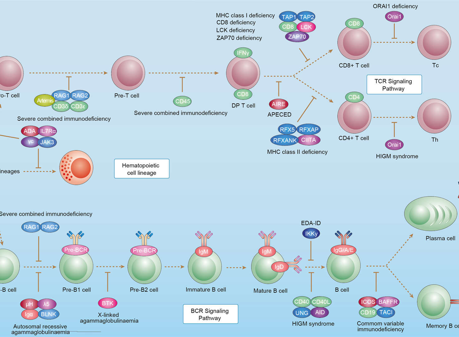 Primary Immunodeficiency
Downloadable Resources
Download resources about recombinant antibody development and antibody engineering to boost your research.
Product Notes
This is a product of Creative Biolabs' Hi-Affi™ recombinant antibody portfolio, which has several benefits including:
• Increased sensitivity
• Confirmed specificity
• High repeatability
• Excellent batch-to-batch consistency
• Sustainable supply
• Animal-free production
See more details about Hi-Affi™ recombinant antibody benefits.
Datasheet
MSDS
COA
Certificate of Analysis LookupTo download a Certificate of Analysis, please enter a lot number in the search box below. Note: Certificate of Analysis not available for kit components.
Lot Number:
See other products for "Clone Leu3a"
- CAT
- Product Name
See other products for "CD4"
Select a product category from the dropdown menu below to view related products.
| CAT | Product Name | Application | Type |
|---|---|---|---|
| MOB-1067z | Mouse Anti-CD4 Recombinant Antibody (clone 34D6) | FC, FuncS, ICC, IF, IP, WB | Mouse IgG2b, κ |
| CAT | Product Name | Application | Type |
|---|---|---|---|
| TAB-039 | Anti-Human CD4 Recombinant Antibody (Zanolimumab) | Neut, ELISA, IF, IP, FuncS, FC, IHC | IgG1 - kappa |
| CAT | Product Name | Application | Type |
|---|---|---|---|
| TAB-260 | Anti-Human CD4 Recombinant Antibody (Tregalizumab) | WB, ELISA, FC, IP, FuncS, IF, Neut | IgG1 - kappa |
| CAT | Product Name | Application | Type |
|---|---|---|---|
| TAB-164 | Anti-Human CD4 Recombinant Antibody (TAB-164) | WB, FuncS, IF, Neut, ELISA, FC, IP | IgG1 - kappa |
| CAT | Product Name | Application | Type |
|---|---|---|---|
| TAB-171 | Anti-Human CD4 Recombinant Antibody (TAB-171) | FuncS, IF, Neut, ELISA, FC, IP, ICC | IgG1 - lambda |
| CAT | Product Name | Application | Type |
|---|---|---|---|
| TAB-732 | Anti-CD4 Recombinant Antibody (Clenoliximab) | IF, IP, Neut, FuncS, ELISA, FC, ICC | IgG4 - lambda |
| CAT | Product Name | Application | Type |
|---|---|---|---|
| TAB-107 | Anti-Human CD4 Recombinant Antibody (TAB-107) | IF, IP, Neut, FuncS, ELISA, FC, ICC | IgG4 - kappa |
| CAT | Product Name | Application | Type |
|---|---|---|---|
| TAB-146 | Anti-Human CD4 Recombinant Antibody (TAB-146) | FuncS, IF, Neut, ELISA, FC, IP, ICC | IgG4 - kappa |
| CAT | Product Name | Application | Type |
|---|---|---|---|
| AGTO-G078E | Anti-CD4 immunotoxin M-T151 (IgG)-PE | Cytotoxicity assay, Function study |
| CAT | Product Name | Application | Type |
|---|---|---|---|
| AGTO-G078R | Anti-CD4 immunotoxin M-T151 (IgG)-RTA | Cytotoxicity assay, Function study |
| CAT | Product Name | Application | Type |
|---|---|---|---|
| PABL-033 | Human Anti-CD4 Recombinant Antibody (clone 17b) | ELISA, Block, FC | Human IgG |
| CAT | Product Name | Application | Type |
|---|---|---|---|
| PABL-034 | Mouse Anti-CD4 Recombinant Antibody (PABL-034) | Neut, ELISA, FuncS | Mouse IgG |
| CAT | Product Name | Application | Type |
|---|---|---|---|
| PABL-035 | Mouse Anti-CD4 Recombinant Antibody (clone Q425) | WB, ELISA, FuncS | Mouse IgG, κ |
| CAT | Product Name | Application | Type |
|---|---|---|---|
| PSBL-033 | Human Anti-CD4 Recombinant Antibody (clone 17b); scFv Fragment | WB, ELISA, FuncS | Human scFv |
| CAT | Product Name | Application | Type |
|---|---|---|---|
| PSBL-034 | Mouse Anti-CD4 Recombinant Antibody scFv Fragment (PSBL-034) | Neut, ELISA, FuncS | Mouse scFv |
| CAT | Product Name | Application | Type |
|---|---|---|---|
| PSBL-035 | Mouse Anti-CD4 Recombinant Antibody (clone Q425); scFv Fragment | WB, ELISA, FuncS | Mouse scFv |
| CAT | Product Name | Application | Type |
|---|---|---|---|
| PFBL-033 | Human Anti-CD4 Recombinant Antibody (clone 17b); Fab Fragment | WB, ELISA, FuncS | Human Fab |
| CAT | Product Name | Application | Type |
|---|---|---|---|
| PFBL-034 | Mouse Anti-CD4 Recombinant Antibody Fab Fragment (PFBL-034) | Neut, ELISA, FuncS | Mouse Fab |
| CAT | Product Name | Application | Type |
|---|---|---|---|
| PFBL-035 | Mouse Anti-CD4 Recombinant Antibody (clone Q425); Fab Fragment | WB, ELISA, FuncS | Mouse Fab |
| CAT | Product Name | Application | Type |
|---|---|---|---|
| PABL-426 | Human Anti-CD4 Recombinant Antibody (clone 576) | WB, ELISA, FuncS | Human IgG |
| CAT | Product Name | Application | Type |
|---|---|---|---|
| PFBL-423 | Human Anti-CD4 Recombinant Antibody (clone 576); Fab Fragment | WB, ELISA, FuncS | Human Fab |
| CAT | Product Name | Application | Type |
|---|---|---|---|
| PFBL-424 | Human Anti-CD4 Recombinant Antibody (clone Hu5A8); Fab Fragment | Block, Neut | Human Fab |
| CAT | Product Name | Application | Type |
|---|---|---|---|
| PSBL-423 | Human Anti-CD4 Recombinant Antibody (clone 576); scFv Fragment | WB, ELISA, FuncS | Human scFv |
| CAT | Product Name | Application | Type |
|---|---|---|---|
| PSBL-424 | Human Anti-CD4 Recombinant Antibody (clone Hu5A8); scFv Fragment | Block, Neut | Human scFv |
| CAT | Product Name | Application | Type |
|---|---|---|---|
| TAB-160LC | Anti-Human CD4 Recombinant Antibody (OKT4A) | ELISA, WB |
| CAT | Product Name | Application | Type |
|---|---|---|---|
| TAB-161LC | Anti-Human CD4 Recombinant Antibody (gOKT4A-4) | ELISA | Humanized antibody |
| CAT | Product Name | Application | Type |
|---|---|---|---|
| TAB-162LC | Anti-Human CD4 Recombinant Antibody | ELISA | Chimeric antibody (mouse/human) |
| CAT | Product Name | Application | Type |
|---|---|---|---|
| TAB-166LC | Anti-Human CD4 Recombinant Antibody (M-T412) | ELISA, Inhib |
| CAT | Product Name | Application | Type |
|---|---|---|---|
| TAB-167LC | Anti-Human CD4 Recombinant Antibody (cM-T412) | ELISA, Inhib | Chimeric antibody (mouse/human) |
| CAT | Product Name | Application | Type |
|---|---|---|---|
| TAB-168LC | Anti-Human CD4 Recombinant Antibody (TRX1) | ELISA | Humanized antibody |
| CAT | Product Name | Application | Type |
|---|---|---|---|
| TAB-169LC | Rat Anti-CD4 Recombinant Antibody (TAB-169LC) | ELISA, FC, SPR | Rat IgG |
| CAT | Product Name | Application | Type |
|---|---|---|---|
| TAB-173LC | Anti-Human CD4 Recombinant Antibody (IgA F425-Alg8) | ELISA, Inhib |
| CAT | Product Name | Application | Type |
|---|---|---|---|
| TAB-174LC | Mouse Anti-CD4 Recombinant Antibody (TAB-174LC) | ELISA, FC, IHC, Neut, FuncS | Mouse IgG1 |
| CAT | Product Name | Application | Type |
|---|---|---|---|
| Gly-025LC | Recombinant Anti-Human CD4 Antibody (Fab glycosylation) | ELISA | Humanized antibody |
| CAT | Product Name | Application | Type |
|---|---|---|---|
| NEUT-351CQ | Mouse Anti-CD4 Recombinant Antibody (clone 10C12) | ELISA, WB, IF, Block | Mouse IgG1 |
| CAT | Product Name | Application | Type |
|---|---|---|---|
| NEUT-352CQ | Mouse Anti-CD4 Recombinant Antibody (clone 13B8.2) | Block, ELISA, FC, IHC-Fr, WB | Mouse IgG1, κ |
| CAT | Product Name | Application | Type |
|---|---|---|---|
| NEUT-353CQ | Mouse Anti-CD4 Recombinant Antibody (clone CBL976) | FC, Neut, IF, IHC | Mouse IgG1, κ |
| CAT | Product Name | Application | Type |
|---|---|---|---|
| NEUT-354CQ | Recombinant Mouse Anti-CD4 Antibody (CT-4) | Neut, FC, IHC, IHC-Fr, IHC-P, IP | IgG1, κ |
| CAT | Product Name | Application | Type |
|---|---|---|---|
| NEUT-355CQ | Mouse Anti-CD4 Recombinant Antibody (clone RPA-T4) | FC, CyTOF, Block, IHC | Mouse IgG1, κ |
| CAT | Product Name | Application | Type |
|---|---|---|---|
| NEUT-356CQ | Mouse Anti-CD4 Recombinant Antibody (clone 3-4F4) | Neut, FC, IHC, IHC-Fr, IP, ICC | Mouse IgG1, κ |
| CAT | Product Name | Application | Type |
|---|---|---|---|
| NEUT-357CQ | Rat Anti-CD4 Recombinant Antibody (clone MAB0810) | Block, Depletion, Stim | Rat IgG2b |
| CAT | Product Name | Application | Type |
|---|---|---|---|
| NEUT-358CQ | Rat Anti-CD4 Recombinant Antibody (clone RM4-5) | FC, CyTOF, Depletion, Block, IHC | Rat IgG2a, κ |
| CAT | Product Name | Application | Type |
|---|---|---|---|
| MOR-0560 | Hi-Affi™ Rabbit Anti-CD4 Recombinant Antibody (clone DS560AB) | IHC-P | Rabbit IgG |
| CAT | Product Name | Application | Type |
|---|---|---|---|
| AFC-TAB-039 | Afuco™ Anti-CD4 ADCC Recombinant Antibody, ADCC Enhanced (AFC-TAB-039) | Neut, ELISA, IF, IP, FuncS, FC | ADCC enhanced antibody |
| CAT | Product Name | Application | Type |
|---|---|---|---|
| AFC-TAB-164 | Afuco™ Anti-CD4 ADCC Recombinant Antibody, ADCC Enhanced (AFC-TAB-164) | FuncS, IF, Neut, ELISA, FC | ADCC enhanced antibody |
| CAT | Product Name | Application | Type |
|---|---|---|---|
| AFC-TAB-260 | Afuco™ Anti-CD4 ADCC Recombinant Antibody, ADCC Enhanced (AFC-TAB-260) | ELISA, FC, IP, FuncS, IF | ADCC enhanced antibody |
| CAT | Product Name | Application | Type |
|---|---|---|---|
| AFC-TAB-171 | Afuco™ Anti-CD4 ADCC Recombinant Antibody, ADCC Enhanced (AFC-TAB-171) | FuncS, IF, Neut, ELISA, FC, IP | ADCC enhanced antibody |
| CAT | Product Name | Application | Type |
|---|---|---|---|
| AFC-TAB-107 | Afuco™ Anti-CD4 ADCC Recombinant Antibody, ADCC Enhanced (AFC-TAB-107) | IF, IP, Neut, FuncS, ELISA, FC | ADCC enhanced antibody |
| CAT | Product Name | Application | Type |
|---|---|---|---|
| VS-0724-YC759 | AbPlus™ Anti-Cd4 Magnetic Beads (VS-0724-YC759) | IP, Protein Purification |
| CAT | Product Name | Application | Type |
|---|---|---|---|
| VS-0924-YC114 | Rat Anti-CD4 Recombinant Antibody Hexamer (VS-0924-YC114), CDC Enhanced | ELISA, FC, CDC | Antibody hexamer |
| CAT | Product Name | Application | Type |
|---|---|---|---|
| VS-0924-YC115 | Human Anti-CD4 Recombinant Antibody Hexamer (VS-0924-YC115), CDC Enhanced | ELISA, FC, ADCC, CDC, SPR | Antibody hexamer |
| CAT | Product Name | Application | Type |
|---|---|---|---|
| VS-0924-YC116 | Mouse Anti-CD4 Recombinant Antibody Hexamer (VS-0924-YC116), CDC Enhanced | WB, ELISA, IF, CDC | Antibody hexamer |
| CAT | Product Name | Application | Type |
|---|---|---|---|
| VS-1024-XY95 | Mouse Anti-NHP CD4 Recombinant Antibody (clone RIV7) | IF, FC | Mouse IgG2a, kappa |
| CAT | Product Name | Application | Type |
|---|---|---|---|
| VS-0225-XY48 | CytoStream™ Mouse Anti-CD4 Recombinant Antibody (clone OKT4) | FC | Mouse IgG2b, kappa |
| CAT | Product Name | Application | Type |
|---|---|---|---|
| VS-0225-XY49 | CytoStream™ Mouse Anti-CD4 Recombinant Antibody (clone OX-35) | FC | Mouse IgG2a, kappa |
| CAT | Product Name | Application | Type |
|---|---|---|---|
| VS-0225-XY50 | CytoStream™ Rat Anti-CD4 Recombinant Antibody (clone RM4-4) | FC | Rat IgG2b, kappa |
| CAT | Product Name | Application | Type |
|---|---|---|---|
| VS-0225-XY51 | CytoStream™ Mouse Anti-CD4 Recombinant Antibody (clone KEN-4) | FC, Block, IHC, IP | Mouse IgG2a, kappa |
| CAT | Product Name | Application | Type |
|---|---|---|---|
| VS-0325-FY64 | Human Anti-CD4 (clone TRX1) scFv-Fc Chimera | ELISA | Human IgG1, scFv-Fc |
| CAT | Product Name | Application | Type |
|---|---|---|---|
| VS-0425-YC755 | Recombinant Anti-CD4 Vesicular Antibody, EV Displayed (VS-0425-YC755) | ELISA, FC, Neut, Cell-uptake |
| CAT | Product Name | Application | Type |
|---|---|---|---|
| VS-0525-XY1178 | Anti-CD4 Immunohistochemistry Kit | IHC |
| CAT | Product Name | Application | Type |
|---|---|---|---|
| VS-0525-XY1179 | Anti-Mouse CD4 Immunohistochemistry Kit | IHC |
| CAT | Product Name | Application | Type |
|---|---|---|---|
| VS-0525-XY1180 | Anti-Human CD4 Immunohistochemistry Kit | IHC |
| CAT | Product Name | Application | Type |
|---|---|---|---|
| VS-0525-YC204 | Recombinant Anti-CD4 (Domain 1 x Domain 2) Biparatopic Antibody, Tandem scFv | ELISA, FC, ICC, IF, IP, Neut | Tandem scFv |
| CAT | Product Name | Application | Type |
|---|---|---|---|
| VS-0825-YC56 | SmartAb™ Recombinant Anti-CD4 pH-dependent Antibody (Clone Zanolimumab) | Neut, ELISA, IF, IP, FC, IHC | Human IgG1 kappa |
| CAT | Product Name | Application | Type |
|---|---|---|---|
| VS-1225-XY51 | Mouse Anti-CD4 Recombinant Antibody (VS-1225-XY51) | FC | Mouse IgG1 |
| CAT | Product Name | Application | Type |
|---|---|---|---|
| VS-1225-XY52 | Mouse Anti-CD4 Recombinant Antibody (VS-1225-XY52) | WB, IHC | Mouse IgG1 |
| CAT | Product Name | Application | Type |
|---|---|---|---|
| VS-1225-XY53 | Mouse Anti-CD4 Recombinant Antibody (VS-1225-XY53) | WB, IHC | Mouse IgG1 |
| CAT | Product Name | Application | Type |
|---|---|---|---|
| VS-1225-XY54 | Mouse Anti-CD4 Recombinant Antibody (VS-1225-XY54) | FC, ELISA | Mouse IgG1 |
| CAT | Product Name | Application | Type |
|---|---|---|---|
| VS-1225-XY55 | Mouse Anti-CD4 Recombinant Antibody (VS-1225-XY55) | IHC, FC, IP | Mouse IgG1 |
Specific Inquiry

See Our Custom Production in Action


Popular Products

Application: WB, IF, IP, Neut, FuncS, ELISA, FC

Application: WB, IP, IF, FuncS, FC, Neut, ELISA

Application: FC, IP, ELISA, Neut, FuncS, IF, WB

Application: WB, FuncS, IF, Neut, ELISA, FC, IP

Application: Inhib, Cyt

Application: IF, IP, Neut, FuncS, ELISA, FC, ICC

Application: IP, IF, FuncS, FC, Neut, ELISA, IHC

Application: FuncS, IF, Neut, ELISA, FC, IP, IHC


Application: ELISA, WB, BLI, SPR


Application: Neut, ELISA, FuncS


For research use only. Not intended for any clinical use. No products from Creative Biolabs may be resold, modified for resale or used to manufacture commercial products without prior written approval from Creative Biolabs.
Send Inquiry
This site is protected by reCAPTCHA and the Google Privacy Policy and Terms of Service apply.