

Activation Assay Protocol & Troubleshooting
Assay protocols for cellular activation provide an effective way to be able to determine immune competence and cellular responsiveness. The interaction between activated immune cells and antigen-presenting cells is an important part of the regulation of the immune response. During the immune response, activated T cells rapidly expand to target and attack tumors. T cells express different activation molecules on the cell surface at different stages of activation, which are criteria for detecting whether T cells are effectively induced to activate.
Creative Biolabs provides a general protocol for T cell activation detection by flow cytometry. The protocol provided here includes the following sections, which you can refer to accordingly to your experimental requirements.
Solutions and Reagents
| Stages | Solutions and Reagents |
| Cell Preparation | Phosphate buffer (PBS), dilution buffer |
| Antibody Activation | CD3 antibody, dilution buffer, washing buffer, CD28 antibody |
Activation Assay Procedure
We describe one of the most common methods to assess T-cell activation. This method measures T cell proliferation after in vitro stimulation of T cells by agonistic antibodies directed against the TCR.
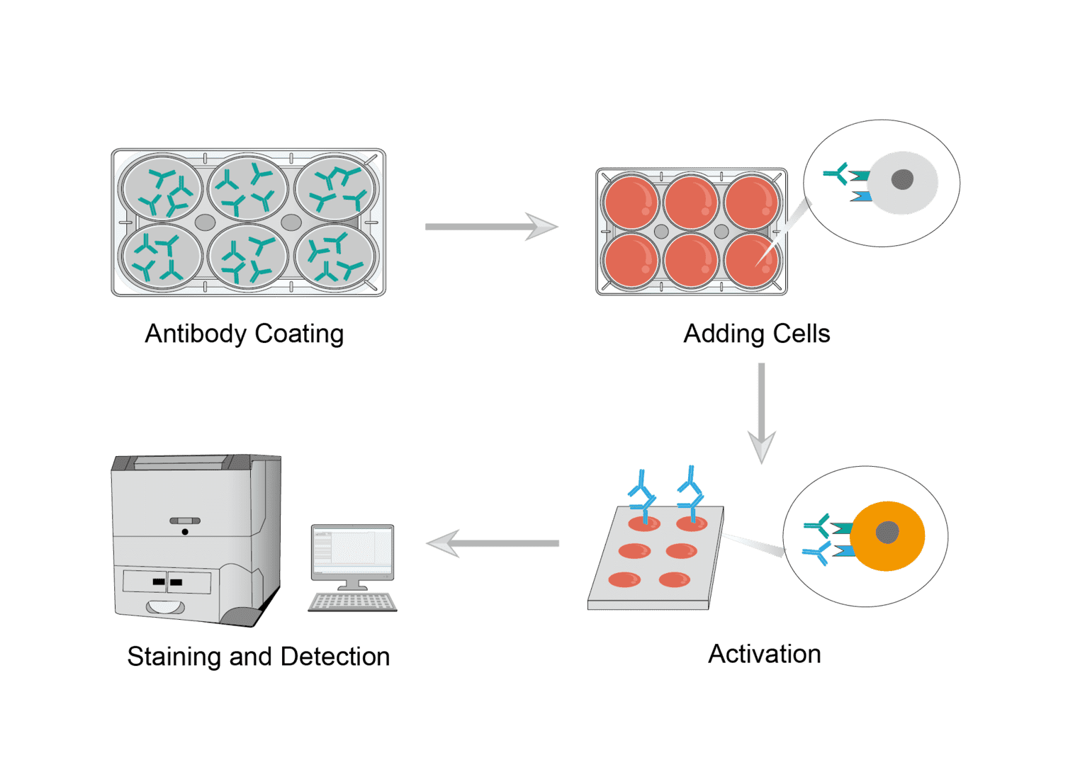
1. Antibody Coating
Prepare the anti-CD3 antibody solution in sterile buffer solution. Then add an equal amount of antibody to each well of a well plate, seal and incubate overnight. Blank controls are incubated with sterile buffer. After incubation, remove the supernatant and wash the plate to remove unbound antibodies.
2. Adding Cells
Collect the cells and resuspend them using buffer. Then centrifuge the resuspended cell samples and remove the supernatant. Repeat several times to obtain a clean sample of cell suspension. Resuspend again and adjust the number of cells. Finally, add an equal number of cells to each well in the well plate.
3. Activation
After the cells are added, add the prepared CD28 antibody dilution solution. And incubate in the incubator for the appropriate time as needed.
4. Staining and Detection
You can perform surface or intracellular staining depending on the cells to be harvested. For more information, you can refer to the flow cytometry protocol. Finally, obtain assay data by flow cytometry.
Troubleshooting
Assay performance is variable
- Inconsistent condition causes. If the target cells were not grown under controlled and consistent conditions, assay performance will vary. We recommend that you follow cell culture guidelines to ensure accurate and consistent cell seeding densities and culture volumes. Monitor cell growth to maintain consistency.
Low measurement
- Instrument causes. Please select an instrument designed for plate reading luminescence detection. We do not recommend using a photometer to measure relative values.
- Insufficient cell causes. Insufficient number of cells per well will result in a low reading. Please follow the instructions for handling and adding cells to ensure a sufficient number of viable cells per well.
- Antibody reagent causes. Low activity of antibody reagents can result in low readings. First you need to store and handle the reagents according to the instructions. Then you can increase the concentration or amount of antibody as appropriate.
Weak assay response
- Sample causes. Please optimize the concentration range of your test sample to achieve a complete dose response with a full upper and lower asymptote.
- Incubation time causes. Optimize the incubation time of the assay in the range of 3-24 hours.
Creative Biolabs is committed to helping you with your experiments for cell activation assays. If you have any requirements, our services can be customized for you.
For research use only. Not intended for any clinical use.
Send Inquiry
This site is protected by reCAPTCHA and the Google Privacy Policy and Terms of Service apply.