

Western Blot Protocol & Troubleshooting
Solutions and Reagents Workflow Troubleshooting Products with Tested Data
Western blot, also known as immunoblot, is a widely used technique for the detection and quantification of specific proteins in complex biological samples. It combines the resolution of gel electrophoresis with the specificity of antibody-based detection. Western blotting is highly versatile and can be used to study protein expression, protein-protein interactions, and post-translational modifications.
Here we go over the western blot protocols, including the reagents, solutions, procedures, and troubleshooting advice for typical issues. Our aim is to help you understand and explain the protocol processes and take full advantage of the detailed protocols for western blot.
Solutions and Reagents
| Stages | Solutions and Reagents |
| Preparation | Lysis buffer, protease inhibitors, phosphate buffer (PBS), sample buffer |
| Electrophoresis | Running buffer, gel solutions, molecular weight markers |
| Transfer | Transfer buffer, methanol, membrane (PVDF or nitrocellulose) |
| Detection | Blocking solution, primary antibody, secondary antibody, washing buffer |
| Visualization | Detection reagents (ECL, chromogenic or fluorescent), developing solution |
Workflow
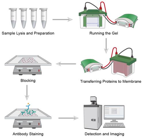 Workflow of Western Blot
1. Sample Preparation
Extract proteins from the sample using appropriate lysis buffer with protease inhibitors. Determine protein concentration using a suitable assay (Bradford, BCA, etc.). Mix samples with sample buffer containing reducing agent and heat at 95°C for denaturation.
2. Gel Electrophoresis
Load protein samples and molecular weight markers onto the gel. Run the gel at appropriate voltage until the dye front reaches the bottom. Ensure proper separation of proteins by monitoring the molecular weight markers.
3. Transfer
Assemble the transfer sandwich with the gel and membrane. Apply current for optimal transfer time based on protein size. Verify transfer efficiency by staining the membrane or gel.
4. Blocking
Incubate the membrane in blocking solution to prevent non-specific binding. Block for the appropriate duration at room temperature with gentle agitation.
5. Antibody Incubation
Incubate with primary antibody at optimized concentration and duration. Wash membrane thoroughly to remove unbound antibody. Incubate with HRP- or fluorophore-conjugated secondary antibody. Perform multiple washing steps.
6. Detection
Apply detection reagents to the membrane according to manufacturer's protocol. Visualize protein bands using appropriate imaging systems. Document and analyze results.
Troubleshooting
Despite the simplicity of the western blot, problems may still arise such as undetected or weak signals. For some of the problems that may occur, we provide the following troubleshooting guide.
- No or Weak Signal
(a) Antibody causes. Primary or secondary antibody concentration may be too low - optimize concentrations through titration; antibody may be incompatible with the detection system - verify antibody-secondary pair; antibodies may have degraded - use fresh antibodies and store properly.
(b) Protein transfer issues. Inefficient transfer to membrane - verify transfer conditions and buffer composition; proteins not retained on membrane - ensure proper membrane activation; large proteins may transfer inefficiently - extend transfer time or use different buffer.
(c) Sample issues. Insufficient protein loading - increase sample concentration or loading volume; target protein may be degraded - use fresh samples and protease inhibitors; protein may not be expressed - verify with positive control.
(d) Detection issues. Reagents may be expired or improperly prepared - use fresh reagents; exposure time too short - optimize exposure duration; substrate concentration incorrect - follow manufacturer's recommendations.
- High Background
(a) Blocking issues. Insufficient blocking - increase blocking time or concentration; incompatible blocking agent - try different blocking solutions; washing inadequate - increase number or duration of washes.
(b) Antibody issues. Antibody concentration too high - optimize through titration; non-specific binding - use more specific antibodies or blocking peptide; cross-reactivity with similar proteins - verify antibody specificity.
(c) Membrane issues. Membrane not properly wetted - ensure complete membrane activation; membrane contamination - handle with clean gloves and forceps; air bubbles during transfer - remove bubbles carefully.
- Multiple or Non-specific Bands
(a) Sample issues. Protein degradation - use protease inhibitors and fresh samples; sample overloading - reduce protein amount; post-translational modifications - consider protein isoforms.
(b) Antibody issues. Non-specific binding - optimize antibody concentration; cross-reactivity - use more specific antibodies; multiple epitope recognition - verify antibody specificity.
- Poor Transfer
(a) Buffer issues. Buffer composition incorrect - verify recipe and pH; methanol concentration improper - adjust for protein size; buffer exhaustion - use fresh buffer.
(b) Technical issues. Air bubbles in transfer sandwich - remove all bubbles; incorrect transfer orientation - verify proper assembly; inadequate power supply - check voltage and current settings.
- Distorted Bands
(a) Gel issues. Sample overloading - reduce protein amount; improper sample preparation - ensure complete denaturation; gel polymerization problems - prepare fresh acrylamide solutions.
(b) Running conditions. Voltage too high - optimize running conditions; temperature effects - use cooling system if necessary; buffer depletion - replace running buffer.
- Uneven Detection
(a) Technical issues. Uneven substrate application - apply carefully and evenly; membrane drying during procedure - keep membrane moist. irregular agitation - ensure consistent shaking.
(b) Equipment issues. Uneven illumination - calibrate imaging system; developer issues - maintain equipment properly; scanner artifacts - clean imaging surface.
Products with Tested Data
At Creative Biolabs, we are dedicated to providing high-quality antibodies for various research applications. Each product in our extensive range has been rigorously tested to ensure superior reliability and efficacy. To showcase the performance of our antibodies, we have conducted numerous experiments using Western Blot. Below, you will find a table listing a selection of our antibody products along with images from these Western Blot experiments, demonstrating their proven reliability.
| Product Name | Catalog Number | Target | Image | Description |
| Humanized Anti-TFRC Recombinant Antibody (TAB-611CT) | HSP90AB1 | TfR |

|
Western blot analysis of TAB-611CT was performed with Recombinant Human Transferrin Receptor Protein. Lane 1: Blank Lane 2: Blank Lane 3: 40ng Recombinant Cynomolgus Transferrin Receptor Protein Lane 4: 40ng Recombinant Cynomolgus Transferrin Receptor Protein Lane 5: Marker Lane 6: 40ng Recombinant Human Transferrin Receptor Protein Lane 7: 80ng Recombinant Human Transferrin Receptor Protein Lane 8: 160ng Recombinant Human Transferrin Receptor Protein Lane 9: 320ng Recombinant Human Transferrin Receptor Protein Lane 10: Blank |
| Anti-PDCD1 Recombinant Antibody (TAB-770) | IκB β | PDCD1 |

|
WB analysis of TAB-770 was performed by with PD-1 Protein, Human, Recombinant (His Tag). Lane 1: TAB-770 antibody Lane 2: Negative control Lane 3: 100ng PDCD1 antigen Lane 4: 200ng PDCD1 antigen Lane 5: 400ng PDCD1 antigen |
| Humanized Anti-TFRC Recombinant Antibody; Fab Fragment (TAB-611CT-F(E)) | H3F3A (Tri Methyl Lys79) | TfR |
-2-1.png)
|
WB analysis of TAB-611CT-F(E) was performed by coating with Transferrin Receptor / TFRC Protein, Human, Recombinant (His Tag). The secondary antibody: HRP-Anti-Human IgG (Fab) Lane 1: Reducing antigen (0.1μg) Lane 2: Reducing antigen (0.3μg) Lane 3: Reducing antigen (0.6μg |
| Anti-Human TIM3 Recombinant Antibody (TSR-022) | HSP70 | TIM-3 |

|
Western blot analysis of TAB-0366CL was performed with Recombinant Human TIM3, His tag in non-reduced (lane 1, 0.5 μg) and reduced condition (lane 2, 0.5 μg) onto a 12% Tris-HCl polyacrylamide gel. Proteins were transferred to a CN membrane and blocked with 5% skim milk for at least one hour. Membranes were probed with TAB-0366CL and HRP Goat Anti-Human IgG as a secondary antibody. Chemiluminescent detection was performed. |
| Mouse Anti-TNFRSF17 Recombinant Antibody (TAB-1223CL) | H3F3A | TNFRSF17 |

|
Western blot analysis of TAB-1223CL (2 μg/mL) was performed with Recombinant Human BCMA Protein (His Tag). The HRP-Anti-Human IgG (H+L) (1: 6000) was used as a secondary antibody. Lane 1: Reducing Antigen (0.1 μg) Lane 2: Reducing Antigen (0.2 μg) Lane 3: Reducing Antigen (0.4 μg) |
| Anti-Human CD2 Recombinant Antibody (TAB-104) | CASP3 | CD2 |

|
Western blot analysis of TAB-104 was performed by loading Recombinant Human CD2 Protein (His Tag) onto a 12% Tris-HCl polyacrylamide gel. Proteins were transferred to a CN membrane and blocked with 5% skim milk for at least one hour. Membranes were probed with TAB-104 and HRP Goat Anti-Human IgG as a secondary antibody. Chemiluminescent detection was performed. Lane 1: Non-Reduced Antigen (1 μg) Lane 2: Reduced Antigen (1 μg) |
| Human Anti-CD47 Recombinant Antibody (HPAB-0008-FY) | LC3A | CD47 |

|
Western blot analysis of HPAB-0008-FY was performed by loading Recombinant Human CD47 Protein (lane 1, non-reducing antigen: 1 μg) onto a 12% Tris-HCl polyacrylamide gel. Proteins were transferred to a CN membrane and blocked with 5% skim milk for at least one hour. Membranes were probed with HPAB-0008-FY and Goat Anti-Human IgG-HRP as a secondary antibody. Chemiluminescent detection was performed. |
| Human Anti-PLAUR Recombinant Antibody (TAB-235CQ) | LTF | PLAUR |

|
Western blot analysis of TAB-235CQ was performed with Recombinant Human PLAUR Protein (His Tag). Lane 1: Non-reducing antigen (0.2μg) Lane 2: Reducing antigen (1μg) The secondary antibody: HRP Goat Anti-Human IgG (H+L) |
| Human Anti-CD19 Recombinant Antibody; scFv Fragment (HPAB-0268-CN-S(P)) | Kif 7 | CD19 |
-2-1.png)
|
Western blot analysis of HPAB-0268-CN-S(P) was performed with CD19 Protein, Human, Recombinant (His Tag). HPAB-0268-CN-S(P)-Biotin incubation concentration: 1.72ng/μL. The secondary antibody: SA-HRP Lane 1: Reducing Antigen (0.1μg) Lane 2: Reducing Antigen (0.3μg) Lane 3: Reducing Antigen (0.6μg) |
| Anti-Human TNFRSF9 Recombinant Antibody (TAB-457CQ) | MKI67 | TNFRSF9 |

|
Western blot analysis of TAB-457CQ was performed with Recombinant Human TNFRSF9 Protein onto a 12% Tris-HCl polyacrylamide gel. Proteins were transferred to a CN membrane and blocked with 5% skim milk for at least one hour. Membranes were probed with TAB-457CQ and HRP Goat Anti-Human IgG as a secondary antibody. Chemiluminescent detection was performed. |
| Human Anti-IL17A Recombinant Antibody (HPAB-0208-LSX) | LMNB1 | IL17A |

|
Western blot analysis of HPAB-0208-LSX (2 μg/mL) was performed with human IL17A (aa 1-155). The HRP-Anti-Human IgG (H+L) (1: 6000) as a secondary antibody. Lane 1: Reducing Antigen (0.1 μg) Lane 2: Reducing Antigen (0.4 μg) Lane 3: Reducing Antigen (0.6 μg) |
| Human Anti-SNAP25 Recombinant Antibody (HPAB-0546-FY) | DDX5 | SNAP25 |

|
Western blot analysis of HPAB-0546-FY was performed with Human SNAP25 Protein (His Tag). |
| Human Anti-CD19 Recombinant Antibody (HPAB-0421-WJ) | TP53 | CD19 |

|
Western blot analysis of HPAB-0421-WJ was performed with Recombinant Human CD19 Protein onto a 12% Tris-HCl polyacrylamide gel. Proteins were transferred to a CN membrane and blocked with 5% skim milk for at least one hour. Membranes were probed with HPAB-0421-WJ and HRP Anti-Human IgG was used as a secondary antibody. Chemiluminescent detection was performed. |
| Anti-Human CD22 Recombinant Antibody (TAB-176) | PRKDC | CD22 |

|
Western blot analysis of TAB-176 was performed with Recombinant Human CD22 Protein onto a 12% Tris-HCl polyacrylamide gel. Proteins were transferred to a CN membrane and blocked with 5% skim milk for at least one hour. Membranes were probed with TAB-176 and HRP Anti-Human IgG as a secondary antibody. Chemiluminescent detection was performed. |
| Anti-Human CD24 Recombinant Antibody | RAF1 | CD24 |

|
Western blot analysis was performed by loading recombinant human CD24 (RPB143Hu01) onto a 12% Tris-HCl polyacrylamide gel. Proteins were transferred to a CN membrane and blocked with 5% skim milk for at least 1 hour. Membranes were probed with Anti-Human CD24 Antibody (TAB-113CL) at a dilution of 1:1000 overnight at 4°C on a rocking platform. Then probed with Goat anti-Human IgG HRP secondary antibody at a dilution of 1:8,000 for one hour. Chemiluminescent was detected. Lane 1: Non-Reducing Antigen (0.05 μg) Lane 2: Reducing Antigen (0.05 μg) |
| Anti-Human CD28 Recombinant Antibody (TGN1412) | Luciferase | CD28 (cluster of differentiation 28) |

|
Western blot analysis of TAB-064LC was performed by loading Recombinant Human CD28 Protein (11524-HCCH) onto a 12% Tris-HCl polyacrylamide gel. Lane 1: Non-Reducing Antigen (0.5 μg) Lane 2: Reducing Antigen (0.5 μg) |
| Mouse Anti-TIM3 Recombinant Antibody (TAB-667CT) | MICU1 | TIM3 |

|
Western blot analysis of TAB-667CT was performed with Recombinant Human TIM3 Protein (lane 2, 1 μg) onto a 12% Tris-HCl polyacrylamide gel. Proteins were transferred to a CN membrane and blocked with 5% skim milk for at least one hour. Membranes were probed with TAB-667CT and HRP Goat Anti-Human IgG was used as a secondary antibody. Chemiluminescent detection was performed. |
| Anti-Human MET Recombinant Antibody (TAB-521CL) | ERBB3 | MET |

|
Western blot analysis of TAB-521CL was performed with Recombinant Human MET Protein Lane 1: Non-Reducing Antigen (0.5μg) Lane 2: Reducing Antigen (0.5μg) |
| Mouse Anti-PCNA Recombinant Antibody (HPAB-N0376-YC) | RPS27 | PCNA |

|
Western blot analysis of HPAB-N0376-YC was performed with Human PCNA Protein (His Tag) in reduced condition Lane 1, Lane 2 and Lane 3 (0.1 μg, 0.3 μg and 0.6 μg respectively) onto a 12% Tris-HCl polyacrylamide gel. Proteins were transferred to a CN membrane and blocked with 5% skim milk for at least one hour. Membranes were probed with HPAB-N0376-YC (2 μg/mL) and HRP-conjugated goat anti-mouse IgG as a secondary antibody (1:6000). Chemiluminescent detection was performed. |
| Anti-Human Abeta Recombinant Antibody (TAB-229) | ATG5 | Abeta |

|
Western blot analysis was performed by loading 1 µg (lane 1, Non-Reduced) and 1 µg (lane 2, Reduced) recombinant human Beta-amyloid 42 with His & GST Tag (10703-H20E2) onto a 12% Tris-HCl polyacrylamide gel. Proteins were transferred to a CN membrane and blocked with 5% skim milk for at least 1 hour. Membranes were probed with Anti-Human Abeta Antibody (TAB-229) at a concentration of 1 μg/mL overnight at 4°C on a rocking platform. Then probed with Goat anti-Human IgG HRP secondary antibody at a dilution of 1:8,000 for one hour. Chemiluminescent was detected. |
| Anti-SARS-CoV S Recombinant Antibody Fab Fragment (MRO-1214LC-F(E)) | ATG7 | S |
-1-2.png)
|
Western blot analysis of MRO-1214LC-F(E) was performed by loading Human SARS Coronavirus Spike Protein (S1 Subunit, His Tag). MRO-1214LC-F(E) incubation concentration: 5 ng/μL. The secondary antibody: HRP-Anti-Human IgG (H+L) Lane 1: Reducing Antigen (0.5 μg) Lane 2: Reducing Antigen (1 μg) Lane 3: Reducing Antigen (2 μg) |
| Human Anti-DPP4 Recombinant Antibody (HPAB-0215-YC) | RTN3 | DPP4 |

|
Western blot analysis of HPAB-0215-YC (2 μg/mL) was performed with Recombinant Human DPP4/CD26 Protein (His Tag). The HRP-Anti-Human IgG (H+L) (1: 6000) was used as a secondary antibody. Lane 1: Reducing Antigen (0.1 μg) Lane 2: Reducing Antigen (0.3 μg) Lane 3: Reducing Antigen (0.6 μg |
| Anti-SARS-CoV S Recombinant Antibody (SC03-014) | RTN1 | Spike |

|
Western blot analysis of MRO-706LC was performed with Human SARS Coronavirus Spike Protein (S1 Subunit, His Tag). MRO-706LC incubation concentration: 2ng/μL. The secondary antibody: HRP-Anti-Human IgG (H+L) Lane 1: Reducing Antigen (0.1μg) Lane 2: Reducing Antigen (0.3μg) Lane 3: Reducing Antigen (0.6μg) |
| Anti-VEEV E2 Recombinant Antibody (CUF37-2a) | NEFM | E2 protein |

|
Western blot analysis of MRO-1163LC was performed with VEEV E2 glycoprotein. MRO-1163LC incubation concentration: 2ng/μL. The secondary antibody: HRP Goat Anti-Mouse IgG (H+L) Lane 1: Reducing Antigen (0.1μg) Lane 2: Reducing Antigen (0.3μg) Lane 3: Reducing Antigen (0.6μg) |
| Anti-Human F3 Recombinant Antibody (TAB-473CQ) | VIM | F3 |

|
Western blot analysis of TAB-473CQ was performed by loading human F3 protein in reduced condition Lane 1, Lane 2 and Lane 3 (0.1 μg, 0.3 μg and 0.6 μg respectively) onto a 12% Tris-HCl polyacrylamide gel. Proteins were transferred to a CN membrane and blocked with 5% skim milk for at least one hour. Membranes were probed with TAB-473CQ (2 μg/mL) and HRP-conjugated goat anti-human IgG as a secondary antibody (1: 5000). Chemiluminescent detection was performed. |
| Human Anti-TNFRSF8 Recombinant Antibody (HPAB-0330-YC) | ACTA2 | TNFRSF8 |

|
Western blot analysis of HPAB-0330-YC was performed with Recombinant Human TNFRSF8 Protein (His Tag). Lane 1: Non-Reducing antigen (0.5μg) Lane 2: Reducing antigen (0.5μg) The secondary antibody: HRP Goat Anti-Human IgG (H+L) |
| Mouse Anti-CD24 Recombinant Antibody (TAB-006LC) | ENO2 | CD24 |

|
Western blot analysis of TAB-006LC was performed with Recombinant Human CD24, His tag in non-reduced (lane 1, 0.5 μg) and reduced condition (lane 2, 0.5 μg) onto a 12% Tris-HCl polyacrylamide gel. Proteins were transferred to a CN membrane and blocked with 5% skim milk for at least one hour. Membranes were probed with TAB-006LC and HRP Goat Anti-Human IgG was used as a secondary antibody. Chemiluminescent detection was performed. |
| Human Anti-FLT3 Recombinant Antibody (clone NC7) | MAP2 | FLT3 |

|
Western blot analysis was performed by loading 1 µg (lane 1) and 1 µg (lane 2) recombinant human FLT3 antigen onto a 12% Tris-HCl polyacrylamide gel. Proteins were transferred to a CN membrane and blocked with 5% skim milk for at least 1 hour. Membranes were probed with TLT3 antibody (HPAB-0183CQ) at a dilution of 1:1000 overnight at 4°C on a rocking platform. Then probed with Goat anti-Human IgG HRP secondary antibody at a dilution of 1:8,000 for one hour. Chemiluminescent was detected. |
| Human Anti-TREM2 Recombinant Antibody (TAB-169CQ) | α skeletal muscle actin | TREM2 |

|
Lane 1: Non-Reducing Antigen (0.5ug) Lane 2: Reducing Antigen (0.5ug) |
| Anti-Human IL1Beta Recombinant Antibody (Canakinumab) | CASP8 | IL1 beta |

|
Western blot analysis of TAB-021 was performed with Recombinant Human IL1 Beta. Lane 1: Non-Reducing Antigen (1ug) Lane 2: Reducing Antigen (1ug) |
| Mouse Anti-IL1RL1 Recombinant Antibody (TAB-222CT) | KRT8 | IL1RL1 |

|
Western blot analysis of TAB-222CT was performed by loading Mouse IL1RL1 Protein (His Tag) in reduced condition Lane 1, Lane 2, Lane 3 (0.1 μg, 0.3 μg, 0.6 μg) onto a 12% Tris-HCl polyacrylamide gel. Proteins were transferred to a CN membrane and blocked with 5% skim milk for at least one hour. Membranes were probed with TAB-222CT (2 μg/mL) and HRP-conjugated goat anti-mouse IgG as a secondary antibody (1: 6000). Chemiluminescent detection was performed. |
| Anti-Human GPC3 Recombinant Antibody (Codrituzumab) | XRCC4 | GPC3 |

|
Western blot analysis of TAB-H14 was performed by loading Human GPC3 Protein (His Tag) in reduced condition Lane 1 and Lane 2 (20 ng and 50 ng) onto a 12% Tris-HCl polyacrylamide gel. Proteins were transferred to a CN membrane and blocked with 5% skim milk for at least one hour. Membranes were probed with TAB-H14 (2 μg/mL) and HRP-conjugated goat anti-human IgG as a secondary antibody (1: 6000). Chemiluminescent detection was performed. |
| Mouse Anti-PlGF Recombinant Antibody (clone AB047) | α-SMA | PlGF |

|
Western blot analysis of HPAB-0723-FY was performed by loading human PIGF protein in reduced condition Lane 1, Lane 2 and Lane 3 (0.1 μg, 0.3 μg and 0.6 μg respectively) onto a 12% Tris-HCl polyacrylamide gel. Proteins were transferred to a CN membrane and blocked with 5% skim milk for at least one hour. Membranes were probed with HPAB-0723-FY (2 μg/mL) and HRP-conjugated goat anti-mouse IgG as a secondary antibody (1: 5000). Chemiluminescent detection was performed. |
| Human Anti-C5 Recombinant Antibody (PABC-462) | MyHCs | C5 |

|
Western blot analysis of PABC-462 was performed with Recombinant Human C5 Protein (His tag) onto a 12% Tris-HCl polyacrylamide gel. Proteins were transferred to a CN membrane and blocked with 5% skim milk for at least one hour. Membranes were probed with PABC-462 (2 μg/mL) and HRP Goat Anti-human IgG (H&L) as a secondary antibody. Chemiluminescent detection was performed. Lane 1: Reducing Antigen (0.1 μg) Lane 2: Reducing Antigen (0.3 μg) Lane 3: Reducing Antigen (0.6 μg) |
| Mouse Anti-CA9 Recombinant Antibody (clone M75) | EGFR | CA9 |

|
Western blot analysis of PABZ-017 was performed with Human CA9 Protein (His Tag). |
| Anti-Human EFNB2 Recombinant Antibody scFv Fragment (B11) | DES | EphB2 (Ephrin-b2) |
-2-1.png)
|
Western blot analysis of TAB-390MZ-S(P) was performed by with Recombinant Human Ephrin-B2 Fc Chimera Protein. TAB-390MZ-S(P) incubation concentration: 1.25ng/μL. The secondary antibody: HRP-Anti-His tag Lane 1: Reducing Antigen (0.1μg) Lane 2: Reducing Antigen (0.3μg) Lane 3: Reducing Antigen (0.6μg) |
| Anti-Human CD23 Recombinant Antibody (Gomiliximab) | HAO1 | CD23 |

|
Western blot analysis of TAB-231 was performed with Recombinant Human CD23 Protein onto a 12% Tris-HCl polyacrylamide gel. Proteins were transferred to a CN membrane and blocked with 5% skim milk for at least one hour. Membranes were probed with TAB-231 and HRP Anti-Human IgG as a secondary antibody. Chemiluminescent detection was performed. |
| Human Anti-SIRPA Recombinant Antibody (TAB-453CT) | TUBE1 | SIRPA |

|
Western blot analysis of TAB-453CT was performed by loading Human SIRPA Protein (His Tag) in reduced condition Lane 1, Lane 2, Lane 3 (0.1 μg 0.3 μg 0.6 μg) onto a 12% Tris-HCl polyacrylamide gel. Proteins were transferred to a CN membrane and blocked with 5% skim milk for at least one hour. Membranes were probed with TAB-453CT (2 μg/mL) and HRP-conjugated goat anti-human IgG as a secondary antibody (1: 6000). Chemiluminescent detection was performed. |
| Human Anti-DR5 Recombinant Antibody (clone Apomab) | MAPK3 | DR5 |

|
Western blot analysis of PABL-453 was performed by loading Recombinant Human DR5 Protein (his Tag) onto a 12% Tris-HCl polyacrylamide gel. Proteins were transferred to a CN membrane and blocked with 5% skim milk for at least one hour. Membranes were probed with PABL-453 and HRP Goat Anti-Human IgG as a secondary antibody. Chemiluminescent detection was performed. |
| Mouse Anti-SNCA Recombinant Antibody (TAB-0750CLV) | LGALS3 | Alpha-synuclein |

|
Western blot analysis was performed by loading Recombinant human alpha-Synuclein Protein (12093-HNAE) onto a 12% Tris-HCl polyacrylamide gel. Proteins were transferred to a CN membrane and blocked with 5% skim milk for at least 1 hour. Membranes were probed with Anti-Human SNCA Antibody (Syn-O4) (TAB-0750CLV) at a dilution of 1:1000 overnight at 4°C on a rocking platform. Then probed with Goat anti-mouse IgG HRP secondary antibody at a dilution of 1:8,000 for one hour. Chemiluminescent was detected. Lane 1: Non-Reducing Antigen (2.5ug) Lane 2: Reducing Antigen (2.5ug) |
| Recombinant Human Anti-RSV Antibody (D25) | NBR1 | RSV |

|
Western blot analysis was performed with Human respiratory syncytial virus (RSV) (A2) Fusion glycoprotein / RSV-F Protein (His Tag) (11049-V08B) onto a 12% Tris-HCl polyacrylamide gel. Proteins were transferred to a CN membrane and blocked with 5% skim milk for at least 1 hour. Membranes were probed with Anti-RSV Antibody (D25) (PABL-322) at a dilution of 1:1000 overnight at 4°C on a rocking platform. Then probed with Goat anti-Human IgG HRP secondary antibody at a dilution of 1:8,000 for one hour. Chemiluminescent was detected. Lane 1: Reducing antigen (0.1 μg) Lane 2: Reducing antigen (0.3 μg) Lane 3: Reducing antigen (0.6 μg) |
| Anti-Human TNFRSF4 Recombinant Antibody (Vonlerolizumab) | GAPDH | TNFRSF4 |

|
Lane 1: Non-Reducing Antigen (0.5ug) Lane 2: Reducing Antigen (0.5ug) |
| Anti-CD33 Recombinant Antibody (TAB-756) | HIST1H2BB | CD33 |

|
Western blot analysis of TAB-756 was performed by loading Recombinant Human CD33 Protein onto a 12% Tris-HCl polyacrylamide gel. The secondary antibody: Goat Anti-Human IgG-HRP. Lane 1: Non-Reducing Antigen (0.5 μg) Lane 2: Reducing Antigen (0.5 μg) |
| Recombinant Anti-human LY6E Antibody | RELA | LY6E |

|
Western blot analysis of MOB-636 was performed by loading human LY6E protein (Lane 1: 0.1 μg, Lane 2: 0.3 μg, Lane 3: 0.6 μg) onto a 12% Tris-HCl polyacrylamide gel. Proteins were transferred to a CN membrane and blocked with 5% skim milk for at least one hour. Membranes were probed with MOB-636 and HRP goat anti-Mouse IgG as a secondary antibody (1: 3000). Chemiluminescent detection was performed. |
| Anti-SARS-CoV S Recombinant Antibody (S230.15) | ERBB2 | Spike |

|
Western blot analysis of MRO-691LC was performed with Human SARS Coronavirus Spike Protein (S1 Subunit, His Tag). MRO-691LC incubation concentration: 2ng/μL. The secondary antibody: HRP-Anti-Human IgG (H+L) Lane 1: Non-Reducing Antigen (0.5μg) Lane 2: Reducing Antigen (0.5μg) |
| Human Anti-TNFRSF10B Recombinant Antibody (TAB-203) | LacZ | TNFRSF10B |

|
Western blot analysis of TAB-203 was performed with DR5/TRAIL R2 Protein, Human, Recombinant (His Tag). TAB-203 incubation concentration: 2ng/μL. The secondary antibody: HRP-Anti-Human IgG (H+L) Lane 1: Reducing Antigen (0.1μg) Lane 2: Reducing Antigen (0.3μg) Lane 3: Reducing Antigen (0.6μg) |
| Human Anti-EPCAM Recombinant Antibody (clone HuMAB UBS-54) | LAMC1 | EPCAM |

|
Western blot analysis of HPAB-0163CQ was performed by loading Recombinant Human EpCAM Protein. Lane 1: Non-Reducing Antigen (1ug). Lane 2: Reducing Antigen (1ug). |
| Mouse Anti-UPAR Recombinant Antibody (clone ATN615) | Cardiotin | UPAR |

|
Western blot analysis of PABC-130 was performed by loading Recombinant Human UPAR Protein (His Tag). Lane 1: Reducing antigen (0.1μg) Lane 2: Reducing antigen (0.5μg) Lane 3: Reducing antigen (1μg) The secondary antibody: HRP Goat Anti-Human IgG (H+L) |
| Anti-human IL23A Recombinant Antibody (TAB-620LC) | RPA2 | IL-23A (Interleukin 23 p19 subunit) |

|
Western blot analysis of TAB-620LC was performed by loading human IL23A protein in reduced condition Lane 1, Lane 2 and Lane 3 (0.1 μg, 0.3 μg and 0.6 μg respectively) onto a 12% Tris-HCl polyacrylamide gel. Proteins were transferred to a CN membrane and blocked with 5% skim milk for at least one hour. Membranes were probed with TAB-620LC (2 μg/mL) and HRP-conjugated goat anti-human IgG as a secondary antibody (1: 5000). Chemiluminescent detection was performed. |
| Human Anti-MSLN Recombinant Antibody (HPAB-0646-CN) | SKP2 | Mesothelin |

|
Western blot analysis of HPAB-0646-CN was performed by loading Mesothelin Protein, Human, Recombinant (His Tag). HPAB-0646-CN incubation concentration: 2 ng/μL. The secondary antibody: HRP-Anti-Human IgG (H+L) Lane: Reducing antigen (0.3 μg) |
References
- Rasheed S, et al. Detailed Western Blotting (Immunoblotting) Protocol. 2022.
- Mahmood T and Yang PC. Western blot: technique, theory, and trouble shooting. N Am J Med Sci, 2012; 4(9): 429-434.
For research use only. Not intended for any clinical use.
Send Inquiry
This site is protected by reCAPTCHA and the Google Privacy Policy and Terms of Service apply.