Bioimaging Protocol & Troubleshooting
Bioimaging is a research tool for understanding various physiological processes in biological cells through the analysis of microstructural images. It includes optical imaging, fluorescence imaging, magnetic resonance imaging, microscopic imaging, molecular imaging, photoacoustic imaging, and ultrasound imaging. With the continuous development of technology, bioimaging has become an indispensable method in cell biology research.
Creative Biolabs offers bioimaging equipment and common protocols with specific service options that can be designed flexibly. We are committed to meeting the technical support for the entire process from experimental design to analysis of results.
Solutions and Reagents
| Stages | Solutions and Reagents |
| Sample Preparation | Phosphate buffer (PBS), culture medium, fixative solution, antibody solution, dye, fluorescein, biotin, probe, washing buffer, mounting solution |
Bioimaging Procedure
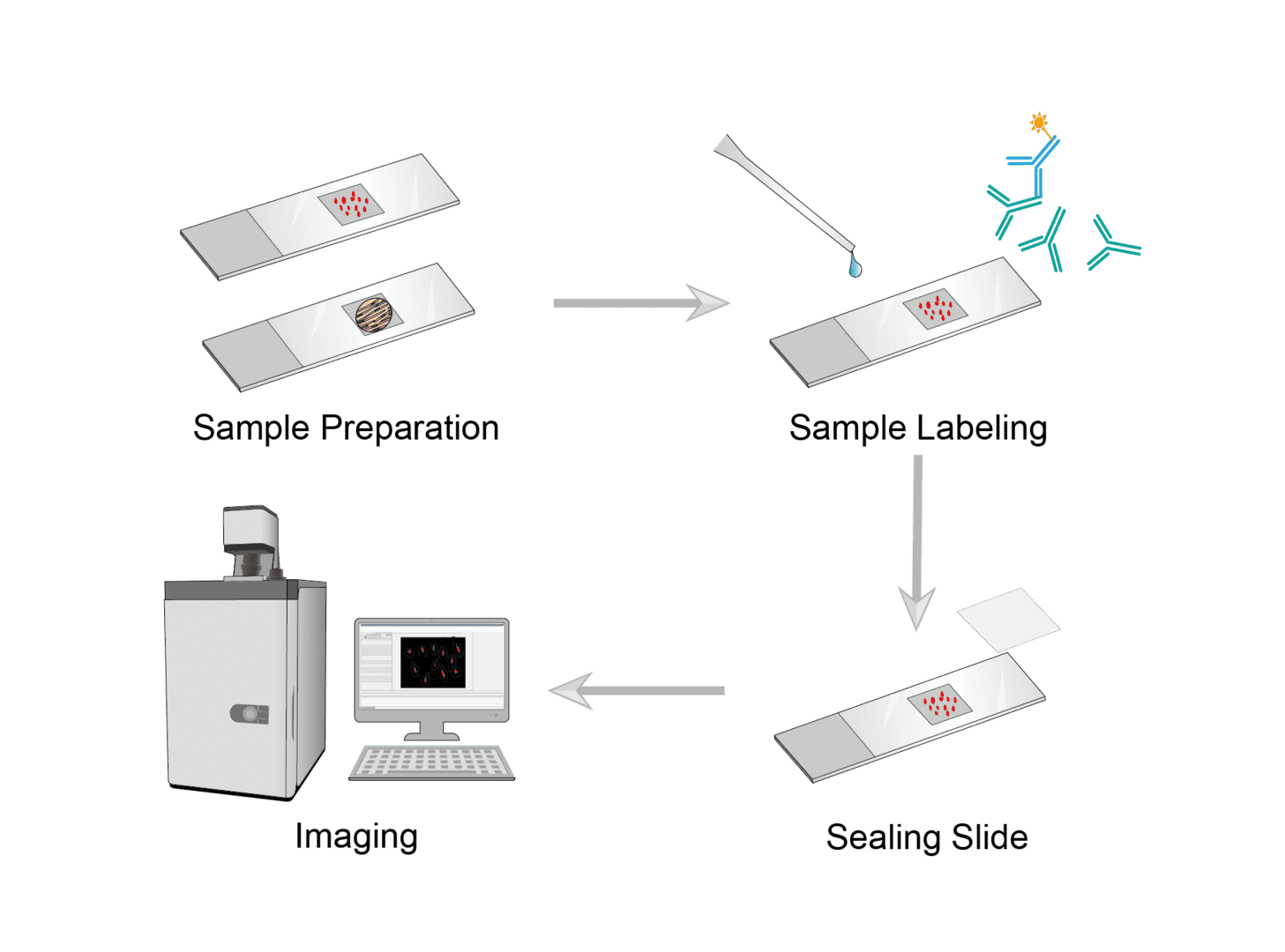
Optical imaging uses light to detect cellular and molecular functions in living organisms. We describe the approximate experimental procedure using optical imaging as an example.

There are different ways of sample preparation for different samples such as cells, tissues, etc. Cell samples are prepared as cell suspensions, added to slides and fixed. Tissue samples can be made into paraffin sections, frozen sections, etc. depending on the specific protocol. The general principle is to make small, thin, intact, transparent samples that retain their original structure.
You can choose from common labeling methods, including staining, immunohistochemistry, in situ hybridization, etc. Common dyes such as hematoxylin, eosin, toluidine blue, etc. are used to stain according to the needs. You can also use fluorescein, biotin and other labeled antibodies to label the corresponding antigens within the sample. And use in situ hybridization to display the nucleic acids to be tested.
Clean the microscope slides and label the slides for each sample. Then add the appropriate amount of mounting medium. Finally place the coverslip and cover it slowly from one side to prevent air bubbles.
After mounting, the sample can be imaged for observation. Substances or physiological processes of interest can be observed by optical imaging instruments such as fluorescence microscopy or confocal microscopy.
Troubleshooting
We summarize some common bioimaging failures and the corresponding solutions. We hope this information can help you to improve the success rate of imaging.
Low signal
- Labeling causes. The possible reason is the unsuitable dyes, fluorescein, biotin, probes. You can reselect a brighter marker.
- Sample mounting causes. The sample is mounted in the incorrect position on the imaging instrument. We recommend using clean coverslips that are sterilized prior to use. Mount the sample as close as possible to the distal end of the coverslip.
- Imaging instrument causes. Check the optical instrument settings used. Please use a high numerical aperture, minimally corrected oil-immersion clean objective lens.
High background
- Cell culture media causes. Note that use a phenol red-free medium.
- Labeling causes. To reduce background and improve signal-to-noise ratio, you can use matching dyes, fluorescein, biotin and other markers for good labeling.
- Other causes. For high background caused by external light, you can turn off the room light and let the light enter through the microscope eyepiece. Choose to use a camera with low readout noise.
Our technical team provides imaging systems and software solutions for your bioimaging and data analysis. The protocol above only briefly describes the general steps of optical imaging, you can contact us for communication and optimization for your specific experiments.
For research use only. Not intended for any clinical use.
This site is protected by reCAPTCHA and the Google Privacy Policy and Terms of Service apply.


