Gel Super Shift Assay Protocol & Troubleshooting
Gel super shift assay is a common technique used to study protein-DNA or protein-RNA interactions. It is used to study protein-DNA interactions by electrophoretic analysis. When electrophoresis is performed, the binding of DNA to proteins causes a shift or delay in the rate of DNA migration. If antibodies that recognize proteins are added to this mixture to create larger complexes, then there is a greater shift. This method is then referred to as a super shift assay.
We perform gel super shift assay experiments using a mixture of nucleic acids loaded with radiolabeled nucleic acids, proteins and protein-specific antibodies following a common protocol. By this method, at least one protein in the complex can be directly identified.
Solutions and Reagents
| Stages | Solutions and Reagents |
| Probe Preparation | Oligonucleotides, dyes, buffer |
| Binding Reaction | Nuclear extract, binding buffer, labeled DNA probe, super shift antibody |
| Electrophoresis | Gel |
Gel Super Shift Assay Procedure
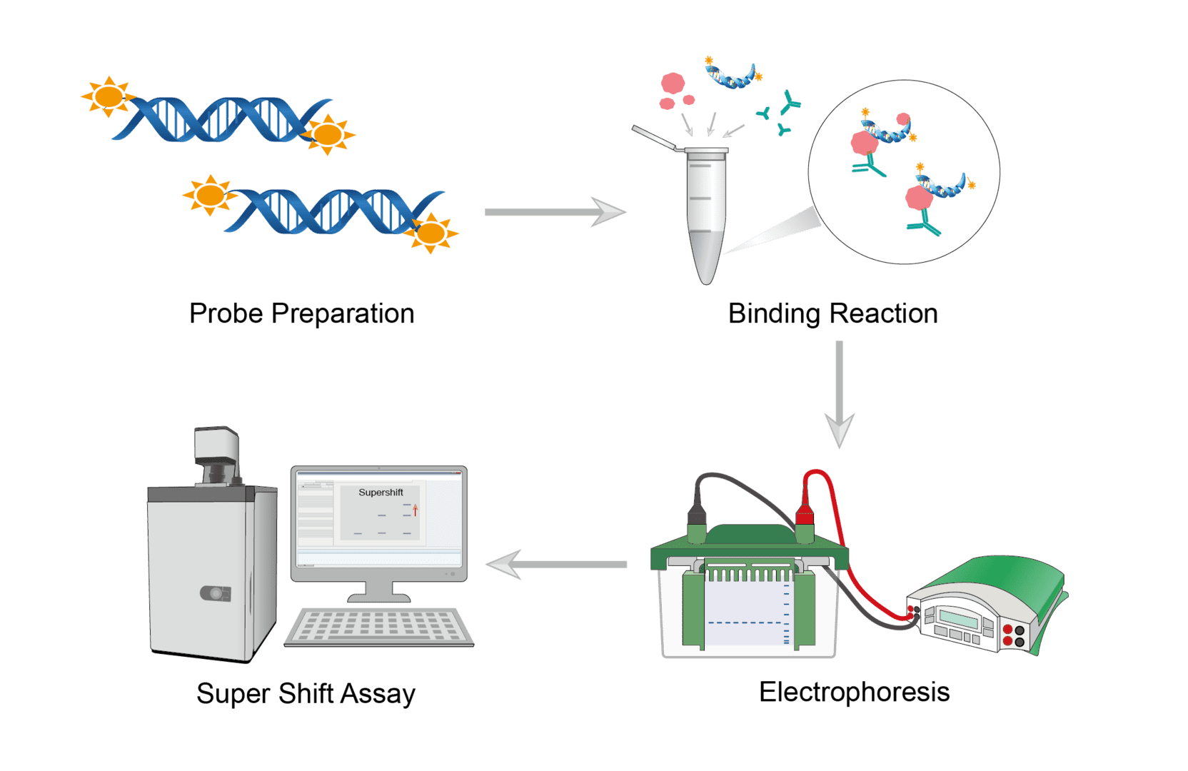
Gel super shift analysis is based on 3 key steps, protein, DNA and antibody binding reaction, electrophoresis and probe detection.

Design and prepare fluorescently labeled or otherwise tagged DNA probes. If the target DNA is short and well-defined, it is possible to synthesize complementary oligonucleotides with specific sequences and anneal them to form double-stranded bodies. If longer DNA fragments are used, it is common to obtain PCR products from plasmids. Then, you can label the DNA by fluorescent staining, chemiluminescence, radioactive methods, etc.
First, add the nuclear extract sample, binding buffer and other reagents to the microtubes and gently mix the contents of the tubes. Then add the specific antibody to the reaction mixture and incubate the reaction at room temperature. Finally, add the labeled probes and mix gently. Incubate the reaction at room temperature.
Prepare the gel. Load all bound reactants onto each lane of the gel and run the gel at the appropriate voltage to the desired distance.
At the end of the electrophoresis run, remove the gel from the electrophoresis unit. Dry the gel and perform a scan to visualize the banding pattern. The bands on the scanned gel can be evaluated using the software.
Troubleshooting
The above protocol provides a reliable framework for gel super shift assay applications. However, there are many issues that can arise when performing specific operations. Below we provide detailed and workable solutions to the relevant common problems.
No protein/oligonucleotide bands on the gel
- Reagent causes. In this case, you should first check your probes and proteins to ensure that no degradation has occurred and that the concentrations are correct. If the above steps do not solve the problem, then you need to make sure that the probe is overloaded according to the amount of protein you are using.
- Binding buffer causes. We recommend changing the composition of the binding buffer and the duration of the binding reaction. Make the time of the binding reaction the first variable to be changed and shorter and longer times should be tested. In addition, you should also change the temperature at which the binding reaction is performed. When making the binding buffer, you should match the endogenous salt concentration of the cell you are interested in as closely as possible to keep the protein stable.
No shift bands detected or weak signal
- Not enough or degraded extract causes. Minimize freezing/thawing cycles of extracts first. Use protease inhibitors to hinder the degradation of the protein. Or add more extracts directly to the reaction.
- Disrupted complex causes. DNA-protein complexes are disrupted due to heating or vortexing. We recommend running the gel with a cooled buffer. Do not vortex the binding reaction.
- Improper scanning causes. You may be using an incorrect scan intensity setting or an incorrect focus offset. You can adjust the focus setting and scan again using the correct scan intensity.
Bands appear smeary or streaky on the gel
- Complex dissociation causes. Band trailing typically occurs when protein/nucleic acid complexes dissociate prior to or during gel electrophoresis. The best way to address this problem is to minimize the time it takes to load the sample as well as the total time the gel is run.
- Gel run system causes. The complex may also dissociate due to the excessive heat generated during the gel run. The cooling system of many gel running systems may not work and we recommend wrapping the outside of the gel running system with ice, or running the gel in a freezer. You can also reduce the voltage during the gel run.
Non-specific competitor reaction
- Nonspecific binding of the analyzed protein to the DNA causes. To prevent non-specific binding of the analyzed protein to DNA, add non-specific competing nucleic acids to the binding reaction. We recommend using non-specific, unlabeled oligonucleotides. Optimize binding by adding different concentrations of competitor.
Complexes appear to be stuck in the wells of the gel
-
Gel causes. This problem is particularly pronounced when performing super shift due to the large antibody/protein/oligonucleotide complexes. To solve this problem, you should first replace the agarose gel with a polyacrylamide gel and use a lower percentage of polyacrylamide gels.
Second, this problem can also be caused by unpolymerized liquid acrylamide remaining in the wells after the gel is injected. This can be simply resolved by thoroughly cleaning the wells before loading the sample.
Finally, you should also start running the gel at a low voltage, rising to full voltage once the sample is in the gel.
We offer standard and common protocols for this technology as well as detailed troubleshooting tips and tricks. If you would like to learn about products and services related to gel super shift assay, please do not hesitate to contact us.
For research use only. Not intended for any clinical use.
This site is protected by reCAPTCHA and the Google Privacy Policy and Terms of Service apply.


