

Apoptosis Protocol & Troubleshooting
Apoptosis is the process of cell self-destruction during normal development. It is prevalent in cell types with a limited lifespan. The problem of regulation of apoptosis is thought to be associated with many diseases. Therefore, apoptosis detection has become a key target in many related research areas.
Apoptosis is a multi-step process. A wide range of apoptosis assays are available for early, intermediate and late cellular events. Creative Biolabs offers a wide range of apoptosis assays including Annexin V binding assay, caspase activity assays, mitochondrial membrane potential assays, DNA fragmentation and morphology.
We provide a general protocol for Annexin V binding assays for apoptosis. This provides a rapid and reliable assay for apoptosis. And these protocols and troubleshooting are useful for flow cytometry or fluorescence microscopy to assess apoptosis.
Solutions and Reagents
| Stages | Solutions and Reagents |
| Cell Seeding | Phosphate buffer (PBS), media, binding buffer |
| Reagent Treatment | Apoptosis reagent, Annexin V incubation reagent, binding buffer |
Apoptosis Procedure
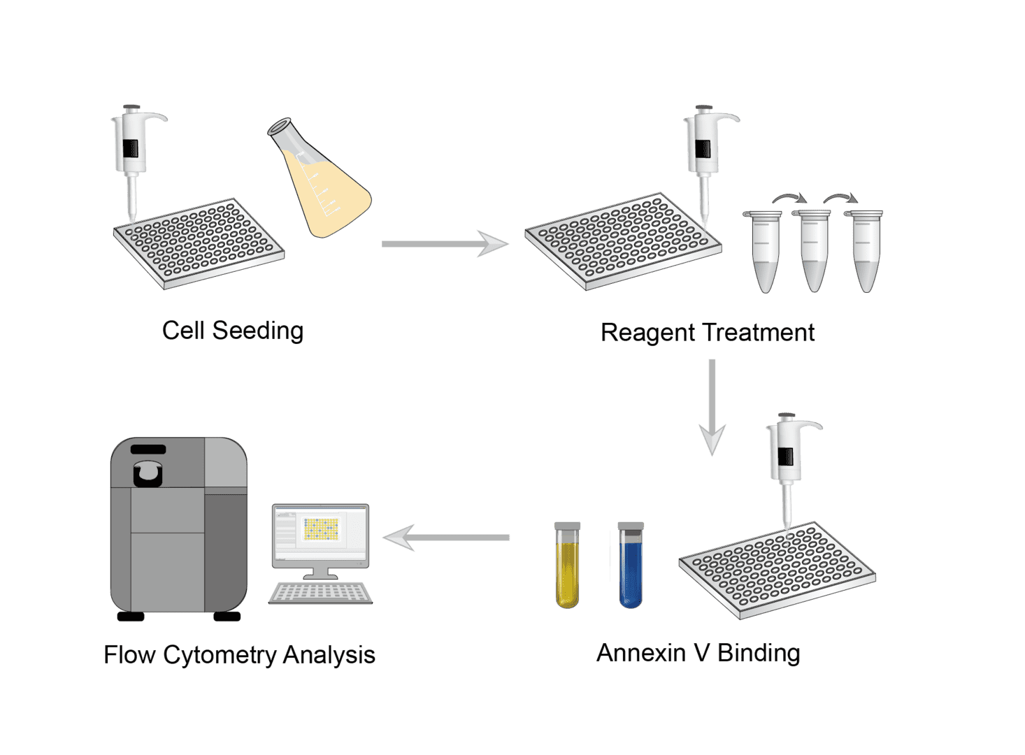
1. Cell Seeding
Collect the cells and wash them. Then dilute the cells to prepare a cell suspension of appropriate concentration. Finally, add the cells and media to a 96-well plate and incubate overnight.
2. Reagent Treatment
Prepare apoptosis reagent. Remove the cell plate from the incubator and aspirate the growth medium. Set up the treatment and control groups in the wells of the 96-well plate, and add the prepared apoptosis reagent respectively.
3. Annexin V Binding
Prepare Annexin V incubation reagent. Add the reagent to the wells and incubate in the dark at room temperature.
4. Flow Cytometry Analysis
Use flow cytometry to analyze cells without washing them.
Troubleshooting
This is a simple experimental protocol. However, there are still some details that need to be paid attention to during the operation. We provide frequently asked questions and their solutions that you can refer to. Please note that you should always use the protocol provided in the data sheet when using a specific kit.
Strong staining in all samples
- Cell damage causes. This can be caused by cells being damaged during harvesting or handling staining. You need to be careful to handle the sample gently during any cell manipulation.
- Unhealthy cell causes. It is possible that your cell sample was unhealthy at the beginning. In this case, you will need to re-run the assay using healthy cells.
No cells seen
- Cells lost causes. If you do not observe cells in the final examination, it is possible that cells have been lost during processing. We recommend that you reduce the processing time or compound dose of the apoptosis reagent.
No signal
- Not enough Annexin V staining solution causes. Not adding enough staining solution can result in low or no signal. You can titrate the optimal staining solution concentration and amount for each type of cell used before the experiment.
- Insufficient stimulation of cell death causes. The stimulus for cell death is not strong enough. You can use a stronger apoptotic stimulus.
Apoptosis is detected by staining cells with Annexin V and propidium iodide solution followed by flow cytometry analysis. This is a simple and rapid method of apoptosis detection. If you need protocols for other apoptosis assays or related products and services, please feel free to contact us.
For research use only. Not intended for any clinical use.
Send Inquiry
This site is protected by reCAPTCHA and the Google Privacy Policy and Terms of Service apply.