

Blocking Protocol & Troubleshooting
Staining antibodies sometimes bind non-specifically to proteins other than the antigen in IHC. To avoid this, blocking experiments can usually be performed. This means that the antibody is neutralized before it is used for staining.
The types of blocking can include two types. The first type is blocking non-specific ion binding. This is due to a number of interactions. In this case, changing the ionic strength of the antibody dilution buffer helps to reduce non-specific ion binding. The second type is blocking endogenous enzymes. When using HRP or AP coupled antibodies for detection, the endogenous level of the enzyme must be blocked.
In general experiments, we typically use serum or bovine serum albumin (BSA) for blocking. Here, we list the blocking procedure and validation protocols for the antibody staining process to help experiments reduce background or non-specific staining.
Solutions and Reagents
| Stages | Solutions and Reagents |
| Blocking | Blocking buffer, antibody solution, blocking peptide |
Blocking Procedure
Blocking reagents are used to effectively reduce the staining background.
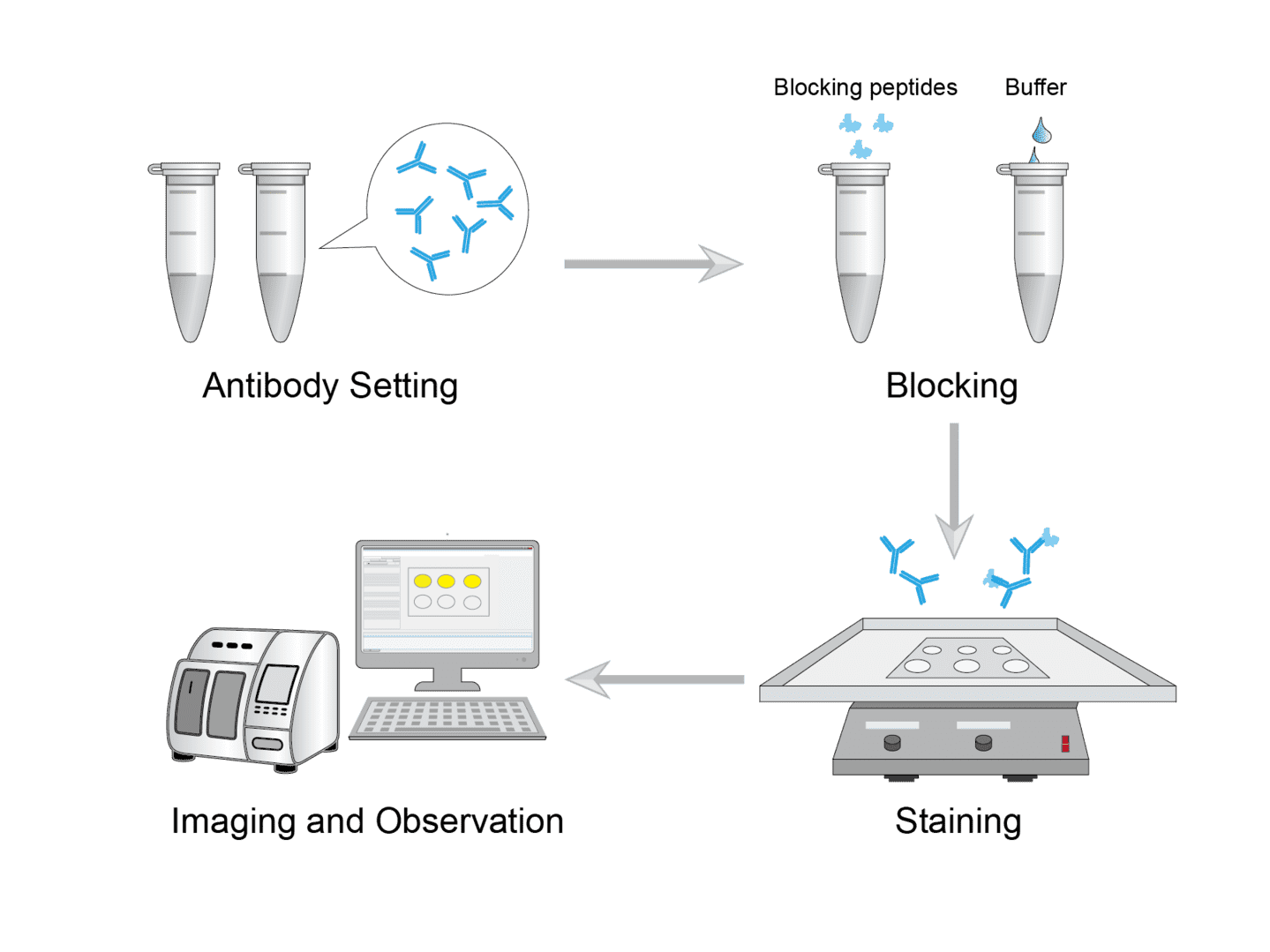
1. Antibody Setting
Determine the type of antibody to be used in your protocol and the optimal antibody concentration. Then determine the amount of antibody needed, and prepare it using the buffer. And set up as two groups, the blocking group and the control group, respectively.
2. Blocking
Add an excess of blocking peptide to the antibody in the blocking group. In the second tube labeled as control, add an equal amount of buffer. Stir both tubes at room temperature and incubate overnight.
3. Staining
Perform the antibody staining protocol on two identical samples. Use blocking antibody for one sample and control antibody for the other sample.
4. Imaging and Observation
Observe the staining results. The staining that disappears when using the blocking antibody is specific to that antibody.
Troubleshooting
Blocking reagents
- Reagent selection. We recommend using serum or bovine serum albumin (BSA) for containment. Serum and BSA help prevent non-specific binding to the many hydrophobic side chains of proteins present in the tissue. If you are using multiple antibodies for staining, you will need to use a blocking serum for all used secondary antibodies.
- Reagent concentration. The concentration of the blocking reagent can be increased by up to 5%. This has a positive effect on reducing background staining. For most standard applications, the recommended concentrations are sufficient. For biotin assays, use a concentration of 5%.
- Reagent setting. When working with negative and positive control samples, select the optimal blocking solution to set the threshold for background staining.
Bocking procedures are often used. Here, we list some tips for reference. You can use blocking buffer to prepare antibody dilutions. As well it is worth noting to ensure that your blocking buffer does not interfere with your signal detection method.
I hope the above information will be helpful to you. If you encounter troubles in your experiments that are not mentioned above, you can contact our technical team for consultation at any time. A well-planned immunization experiment needs to be coupled with proper controls, handling and conditions. This can often help you achieve improved results. For details on how to plan your blocking procedures in specific experiments, please see our other experimental guides.
Products with Tested Data
At Creative Biolabs, we are dedicated to providing high-quality antibodies for various research applications. Each product in our extensive range has been rigorously tested to ensure superior reliability and efficacy. To showcase the performance of our antibodies, we have conducted numerous experiments using the Blocking assay. Below, you will find a table listing a selection of our antibody products along with images from these experiments, demonstrating their proven reliability.
| Product Name | Catalog Number | Target | Image | Description |
|---|---|---|---|---|
| Human Anti-PDCD1 Recombinant Antibody (TAB-H55) | TAB-H55 | PDCD1 |

|
PD-L1 binding to PD-1 was blocked by anti-PD-1 antibody (Cat# TAB-H55), IC₅₀ = 442.9 ng/mL. In a functional ELISA, the recombinant human PD‑1 protein (His tag) was coated at 2 µg/mL, 100 µL/well. Anti-PD-1 antibody (Cat# TAB-H55) were 3-fold serial diluted from 20 µg/mL, 100 µL/well. Coated PD-1 protein was incubated with the human PD-L1 mFc chimera (5 µg/mL) and anti-PD-1 antibody (Cat# TAB-H55). The PD-L1 binding to PD-1 was detected by HRP-goat anti-mouse IgG secondary antibody. Human IgG4 antibody (Cat# MOB-0408ZL, Creative Biolabs) was used as an isotype control. |
| Anti-PDCD1 Recombinant Antibody (TAB-770) | TAB-770 | PDCD1 |

|
PD-L1 binding to PD-1 was blocked by anti-PD-1 antibody (Cat# TAB-770), IC₅₀ = 246.6 ng/mL. In a functional ELISA, the recombinant human PD‑1 protein (His tag) was coated at 2 µg/mL, 100 µL/well. Anti-PD-1 antibody (Cat# TAB-770) were 3-fold serial diluted from 20 µg/mL, 100 µL/well. Coated PD-1 protein was incubated with the human PD-L1 mFc chimera (5 µg/mL) and anti-PD-1 antibody (Cat# TAB-770). The PD-L1 binding to PD-1 was detected by HRP-goat anti-mouse IgG secondary antibody. Human IgG4 antibody (Cat# MOB-0408ZL, Creative Biolabs) was used as an isotype control. |
For research use only. Not intended for any clinical use.
Send Inquiry
This site is protected by reCAPTCHA and the Google Privacy Policy and Terms of Service apply.