

Immunoprecipitation Protocol & Troubleshooting
Immunoprecipitation (IP) is a biotechnology technique for studying protein interactions. This technique enables the isolation and identification of proteins from biological solutions. It is based on the principle of treating proteins as antigens and using antibodies immobilized on solid phase supports such as beads or agarose to specifically bind to them. There are many different types of IP and it is an important step in many proteomic studies.
We briefly overview IP protocols, including reagents used for experiments, buffers, general procedures and troubleshooting tips. By using it in conjunction with immunoblotting or other protein research methods, purified specific proteins can be analyzed or identified.
Solutions and Reagents
| Stages | Solutions and Reagents |
| Sample Preparation | Phosphate buffer (PBS), cell lysis buffer, protease inhibitors, dilution buffer, IP buffer |
| Immunoprecipitation | Antibody, agarose beads, washing buffer, elution buffer |
Immunoprecipitation Procedure
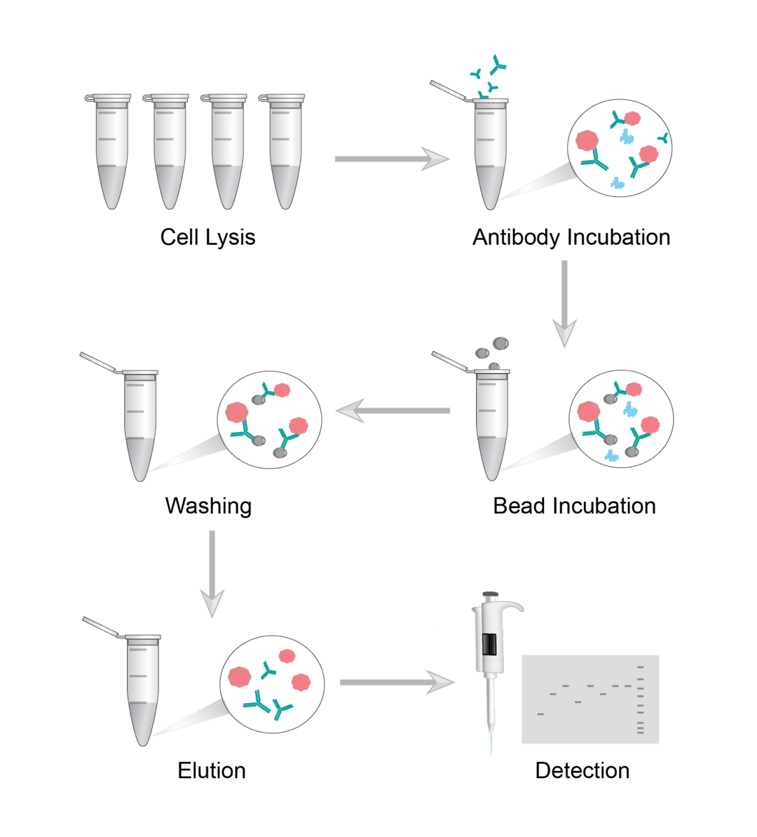
1. Cell Lysis
Scrape the cells from the culture dish into a tube and then centrifuge to collect the cells. Add ice-cold lysis buffer to the cells and sonicate the cells using the appropriate settings. After several repetitions, centrifuge the cells and transfer the supernatant to a new tube. Finally, determine the total protein and adjust the concentration with IP buffer. If tissue samples are used, the tissue is ground using liquid nitrogen and then sonicated. Then prepare the cell lysate as described above.
2. Immunoprecipitation
Dilute the cell lysate. Add the cell lysate and the recommended amount of antibody to a new tube. You may refer to the antibody data sheet for recommended antibody concentrations. Incubate overnight to form an antigen-antibody complex. Add immobilized beads to capture immune complexes. Either agarose beads or magnetic beads may be used. After addition, incubate, centrifuge and carefully and completely remove the supernatant to collect the complex precipitate. If you choose magnetic beads, apply a magnetic field to pull the beads to the side of the tube and carefully pipette the supernatant away. Finally, repeat the wash several times.
3. Washing and Elution
There are various methods that can be used to elute proteins from the beads, such as acid elution and thermal denaturation. Resuspend the beads in buffer and mix gently. Boil at 100 ºC for 5-10 minutes to dissociate the immune complex from the beads. Finally, centrifuge the sample to allow the beads to settle. If you use magnetic beads, a magnetic field is applied to the sample. And carefully collect the supernatant for later use.
4. Detection
Load protein samples onto SDS-PAGE gels and perform western blotting (WB) to detect IP results.
Troubleshooting
Although the IP method is very simple. However, there can be various specific differences between different proteins and different antibodies. Therefore, the variables and factors that affect the success of an experiment are not only numerous and varied. We describe some common troubleshooting tips for you to help you continuously optimize your experimental conditions and finally get satisfactory isolated proteins.
No eluted target protein detected
-
Sample causes. One possibility is that the target protein is not expressed in the sample used or is expressed at a low level. You should check the expression of the target protein. If the target protein expression level is low, you can increase the amount of lysate. But at the same time this may lead to an increase in non-specific binding. Therefore, we recommend pre-clearing the lysate before starting the IP procedure.
Another possibility is that there are too many competing proteins in the sample. We recommend centrifugation after cell lysis to remove insoluble proteins, membrane fragments, debris, etc.
It may also be that the target antigen is lost or destroyed in the sample. You can try preparing fresh lysates under appropriate protease inhibitors and prevent freezing.
Finally, there may also be interfering substances present in the sample. Some lysates containing reducing agents can disrupt antibody function and should be avoided. Extreme pH values and excessive reagent concentrations may also interfere with antibody-antigen interactions. - Lysis buffer causes. It is possible that you used the incorrect lysis buffer. First make sure that you are using the correct lysis buffer. You can use a different lysis buffer to improve solubility. Or try to denature the lysis conditions. On the other hand, the amount of lysis buffer is not enough. You should increase the amount of lysis buffer and pre-clean the lysis buffer before starting the IP procedure.
- Antibody causes. It is possible that you do not have enough antibody to capture the target protein. Check if using the recommended amount of antibody. Determine the optimal concentration of antibody by titration experiments. Or perhaps the antibody you selected is not suitable for immunoprecipitation. We recommend that you try different antibodies and use an antibody with a higher affinity for the target. Polyclonal antibodies usually perform better than monoclonal antibodies. Also, if the primary antibody is not fully captured, use a secondary antibody.
- Bead causes. You may have used the wrong type of beads, resulting in antibodies not binding to the immunosorbent beads. Make sure you are using the correct type of beads. It is also possible that the antibodies are weakly bound to the beads and you can change to another brand of beads.
- Incubation causes. Incubation time is too short. Usually, the antibody and the target antigen are incubated for 4 hours to overnight.
- Washing causes. Too strict washing causes. Reduce the number of washes and detergent concentration. Avoid washing buffer stripping immune complexes from agarose beads.
- Elution causes. The target protein is not eluted from the magnetic beads. You need to make sure that you are using the correct elution buffer and that it has the correct strength and pH.
High background
- Sample causes. When preparing cell lysates, firstly avoid freezing the lysate. Next ensure to add fresh protease inhibitors to prevent antigen degradation. Too many cells can result in a large number of non-specific proteins in the eluate. You can reduce the number of cells/lysates used. As well as reducing the non-specific binding of proteins to beads or antibodies by pre-clearing the lysate if necessary.
- Antibody causes. Too much antibody can lead to non-specific binding. We recommend determining the optimal concentration of antibody by titration. It is also possible that the antibody used is not specific enough, so use an affinity-purified antibody. Precipitation may be present in the antibody and you will need to centrifuge to remove any particulate matter.
- Bead causes. Non-specific protein binding caused by beads that are not adequately pre-closed. Ensure that the blocking agent is fresh and in saturated amounts.
- Washing causes. Washing is not thorough. Be sure to wash well at the relevant stage. Use a more stringent washing solution or increase the number of washes.
Other problems
- A large number of antibodies eluted. We recommend trying to reduce the amount of antibody used. Cross-link the antibody to the beads prior to IP and elute using a mild buffer gradient.
- Protein of interest blocked by antibody heavy or light chains. To avoid interference from the antibody chain, we recommend the use of secondary antibodies.
IP is a well-established technology that has been an important step in many proteomics studies. We are committed to providing you with efficient and simple IP protocols for the successful isolation of sufficient amounts of specific proteins. On our website, you can select the appropriate antibody for IP based on our product information.
Visit our products and services to order assay antibodies and exclusive services.
Products with Tested Data
At Creative Biolabs, we are dedicated to providing high-quality antibodies for various research applications. Each product in our extensive range has been rigorously tested to ensure superior reliability and efficacy. To showcase the performance of our antibodies, we have conducted numerous experiments using Immunoprecipitation (IP) assay. Below, you will find a table listing a selection of our antibody products along with images from these experiments, demonstrating their proven reliability.
| Product Name | Catalog Number | Target | Image | Description |
|---|---|---|---|---|
| Mouse Anti-E-Tag Recombinant Antibody (clone 11H12B3) | ZG-0287U | E-Tag |

|
Immunoprecipitating E-Tag in 293F transfected whole cell lysate Lane 1: Mouse control IgG (1µg) in 293F transfected whole cell lysate. For western blotting, a HRP-conjugated Protein G antibody was used as the secondary antibody (1/2000) Lane 2: ZG-0287U (5µg) + 293F transfected whole cell lysate (500µg) Lane 3: 293F transfected whole cell lysate (20µg) |
| Mouse Anti-ZAP70 Recombinant Antibody (ZG-0515F) | ZG-0515F | ZAP70 |

|
The ZAP-70 mouse mAb was used for immunoprecipitation analysis of Jurkat cell lysates (ZAP-70 positive expressing cell line) and Ramos cell lysates (ZAP-70 negative expressing cell line). |
| Mouse Anti-PODXL Recombinant Antibody (clone 4D2G7) | ZG-0167U | PODXL |

|
Immunoprecipitating PODXL in HEK293 whole cell lysate. Lane 1: Mouse control IgG (1µg) instead of ZG-0167U in HEK293 whole cell lysate. For western blotting, a HRP-conjugated Protein G antibody was used as the secondary antibody (1/2000) Lane 2: ZG-0167U (8µg) + HEK293 whole cell lysate (500µg) Lane 3: HEK293 whole cell lysate (10µg) |
| Mouse Anti-CBX3 Recombinant Antibody (ZG-0443J) | ZG-0443J | CBX3 |

|
Immunoprecipitation analysis of Hela cell lysates using HP1-gamma mouse mAb. |
| Mouse Anti-VIM Recombinant Antibody (ZG-0501F) | ZG-0501F | VIM |

|
Immunoprecipitation analysis of Hela cell lysates using vimentin mouse mAb. |
| Mouse Anti-RAD9 Recombinant Antibody (ZG-052R) | ZG-052R | RAD9 |

|
Immunoprecipitation analysis of Hela and 3T3 cell lysates using RAD9A mouse mAb. |
| Mouse Anti-LMNB1 Recombinant Antibody (clone 7C11) | MOB-0073F | LMNB1 |

|
1) Mouse brain tissue lysate 2) IP product: IP dilution 1:200. |
| Mouse Anti-DLAT Recombinant Antibody (ZG-042R) | ZG-042R | DLAT |

|
Immunoprecipitation analysis of Hela cell lysates using Pyruvate Dehydrogenase E2 mouse mAb. |
| Mouse Anti-IgG Recombinant Antibody (ZG-049R) | ZG-049R | IgG |

|
Immunoprecipitation analysis of cell lysates transfected with HA-tagged NGAL using HA-Tag Rabbit pAb (1:100 diluted) (lane 2). Lane 1 contains rabbit IgG IP control lysate. All lanes are probed with HA-Tag Rabbit pAb (1:1000 diluted) as the primary antibody. Panel A incubated with secondary antibodies Anti-Rabbit whole IgG, HRP-linked antibody for detecting. Panel B incubated with Anti-Rabbit IgG (Heavy-Chain Specific) mAb (1:50000 diluted) and the Anti-mouse whole IgG, HRP-linked antibody was used for detecting. |
| Mouse Anti-RANBP9 Recombinant Antibody (ZG-058R) | ZG-058R | RANBP9 |

|
Immunoprecipitation analysis of Hela cell lysates using RanBP9 mouse mAb. |
| Mouse Anti-SIRT6 Recombinant Antibody (ZG-090R) | ZG-090R | SIRT6 |

|
Immunoprecipitation analysis of Hela cell lysates using SIRT6 mouse mAb. |
| Rabbit Anti-DHFR Recombinant Antibody (clone 9B2) | ZG-0636J | DHFR |

|
Immunoprecipitating DHFR in Hela whole cell lysate Lane 1: Rabbit control IgG in Hela whole cell lysate. For western blotting,a HRP-conjugated Protein G antibody was used as the secondary antibody (1/2000) Lane 2: ZG-0636J (2µg)+ Hela whole cell lysate (500µg) Lane 3: Hela whole cell lysate (10µg) |
| Mouse Anti-HDAC4 Recombinant Antibody (ZG-0417J) | ZG-0417J | HDAC4 |

|
Immunoprecipitation analysis of Hela cell lysates using HDAC4 mouse mAb. |
| Mouse Anti-HSP90AB1 Recombinant Antibody (ZG-0460J) | ZG-0460J | HSP90AB1 |

|
Immunoprecipitation analysis of Hela cell lysates using Hsp90 beta mouse mAb. |
| Mouse Anti-RPA32 Recombinant Antibody (ZG-072R) | ZG-072R | RPA32 |

|
Immunoprecipitation analysis of Hela cell lysates using RPA32/RPA2 mouse mAb. |
| Mouse Anti-RPA70 Recombinant Antibody (ZG-073R) | ZG-073R | RPA70 |

|
Immunoprecipitation analysis of Hela cell lysates using RPA70 mouse mAb. |
| Rabbit Anti-Phospho-POLR2A (S2) Recombinant Antibody (clone 2G1) | ZG-0514U | POLR2A |

|
Immunoprecipitating Phospho-POLR2A in Hela whole cell lysate Lane 1: Rabbit control IgG(1μg)instead of ZG-0514U in Hela whole cell lysate. For western blotting, a HRP-conjugated Protein G antibody was used as the secondary antibody (1/2000) Lane 2: ZG-0514U(3μg)+ Hela whole cell lysate(1mg) Lane 3: Hela whole cell lysate (20μg) |
| Rabbit Anti-PKM Recombinant Antibody (clone 7B2) | ZG-0513U | PKM |

|
Immunoprecipitating PKM in Hela whole cell lysate Lane 1: Rabbit control IgG instead of ZG-0513U in Hela whole cell lysate. For western blotting, a HRP-conjugated Protein G antibody was used as the secondary antibody (1/2000) Lane 2: ZG-0513U(2µg)+ Hela whole cell lysate(500µg) Lane 3: Hela whole cell lysate (10µg) |
| Rabbit Anti-Phospho-RB1 (S780) Recombinant Antibody (clone 2E9) | ZG-0544U | RB1 |

|
Immunoprecipitating Phospho-RB1 in Hela whole cell lysate Lane 1: Rabbit control IgG(1μg)instead of ZG-0544U in Hela whole cell lysate. For western blotting, a HRP-conjugated Protein G antibody was used as the secondary antibody (1/2000) Lane 2: ZG-0544U(3μg)+ Hela whole cell lysate(1mg) Lane 3: Hela whole cell lysate (20μg) |
| Rabbit Anti-Phospho-RPS6KB1 (T421+S424) Recombinant Antibody (clone 3B6) | ZG-0555U | RPS6KB1 |

|
Immunoprecipitating Phospho-RPS6KB1 in Hela whole cell lysate Lane 1: Rabbit control IgG(1μg)instead of ZG-0555U in Hela whole cell lysate. For western blotting, a HRP-conjugated Protein G antibody was used as the secondary antibody (1/2000) Lane 2: ZG-0555U(3μg)+ Hela whole cell lysate(1mg) Lane 3: Hela whole cell lysate (20μg) |
| Mouse Anti-HSPA1A Recombinant Antibody (ZG-0456J) | ZG-0456J | HSPA1A |

|
Immunoprecipitation analysis of Hela cell lysates using Hsp70 (C-terminus) mouse mAb. |
| Rabbit Anti-Phospho-RPS6KA1 (S380) Recombinant Antibody (clone 1E11) | ZG-0553U | RPS6KA1 |

|
Immunoprecipitating Phospho-RPS6KA1 in Hela whole cell lysate Lane 1: Rabbit control IgG(1μg)instead of ZG-0553U in Hela whole cell lysate. For western blotting, a HRP-conjugated Protein G antibody was used as the secondary antibody (1/2000) Lane 2: ZG-0553U(3μg)+ Hela whole cell lysate(1mg) Lane 3: Hela whole cell lysate (20μg) |
| Mouse Anti-ENO1 Recombinant Antibody (clone 4D11F5) | ZG-0251U | ENO1 |

|
Immunoprecipitating ENO1 in HepG2 whole cell lysate Lane 1: Mouse control IgG (1µg) in HepG2 whole cell lysate. For western blotting, a HRP-conjugated Protein G antibody was used as the secondary antibody (1/2000) Lane 2: ZG-0251U (1µl) + HepG2 whole cell lysate (500µg) Lane 3: HepG2 whole cell lysate (10µg) |
| Mouse Anti-PDCD1LG2 Recombinant Antibody (clone 7F11D11) | ZG-0252U | PDCD1LG2 |

|
Immunoprecipitating PD-L2 in Hela whole cell lysate Lane 1: Mouse control IgG in Hela whole cell lysate Lane 2: ZG-0252U (2µl) + Hela whole cell lysate (500µg) Lane 3: Hela whole cell lysate (20µg) For western blotting, the blot was detected with ZG-0252U at 1:2000, and a HRP-conjugated Protein G antibody was used as the secondary antibody at 1:2000 |
| Mouse Anti-SWAP70 Recombinant Antibody (ZG-122R) | ZG-122R | SWAP70 |

|
Immunoprecipitation analysis of Hela cell lysate using SWAP70 mouse mAb. |
| Mouse Anti-IKZF1 Recombinant Antibody (ZG-0485J) | ZG-0485J | IKZF1 |

|
Immunoprecipitation analysis of Hela cell lysate using Ikaros (C-terminus) mouse mAb. |
| Mouse Anti-SHP1 Recombinant Antibody (ZG-086R) | ZG-086R | SHP1 |

|
Immunoprecipitation analysis of Raji cell lysates using SHP-1 mouse mAb. |
| Mouse Anti-SIRT1 Recombinant Antibody (ZG-087R) | ZG-087R | SIRT1 |

|
Immunoprecipitation analysis of Hela cell lysates using SIRT1 mouse mAb. |
| Mouse Anti-STAT1 Recombinant Antibody (ZG-113R) | ZG-113R | STAT1 |

|
Immunoprecipitation analysis of Hela cell lysates using STAT1 mouse mAb. |
| Mouse Anti-ACTA1 Recombinant Antibody (clone 4B11) | ZG-0181C | ACTA1 |

|
1) Mouse Brain tissue Lysate 2) IP product: IP dilute 1: 200 |
| Mouse Anti-XRCC4 Recombinant Antibody (clone 5C10) | ZG-0178C | XRCC4 |

|
1) Hela Cell Lysate 2) IP product: IP dilute 1:200 |
| Mouse Anti-RELA Recombinant Antibody (clone 14H2) | MOB-0143F | RELA |

|
1) Hela Cell Lysate 2) IP products: IP dilution 1:200. |
| Mouse Anti-HSPD1 Recombinant Antibody (ZG-0452J) | ZG-0452J | HSPD1 |

|
Immunoprecipitation analysis of Hela cell lysates using Hsp60 mouse mAb. |
| Mouse Anti-LacZ Recombinant Antibody (clone 3B7D5) | ZG-0244U | LacZ |

|
Immunoprecipitating lacZ in 293T transfected whole cell lysate Lane 1: Mouse control IgG in 293T transfected whole cell lysate Lane 2: ZG-0244U (2µl) + 293T transfected whole cell lysate (500µg) Lane 3: 293T transfected whole cell lysate (20µg) For western blotting, the blot was detected with ZG-0244U at 1:2000, and a HRP-conjugated Protein G antibody was used as the secondary antibody at 1:2000 |
| Mouse Anti-HistoneH3 Recombinant Antibody (clone 1G2E4) | ZG-0277U | HistoneH3 |

|
Immunoprecipitating HistoneH3 in Hela whole cell lysate Lane 1: Mouse control IgG2b in Hela whole cell lysate. Lane 2: ZG-0277U (2μg) + Hela whole cell lysate (500μg) Lane 3: Hela whole cell lysate (20μg) For western blotting, the blot was detected with ZG-0277U at 1:10000, and a HRP-conjugated Protein G antibody was used as the secondary antibody at 1:2000 |
| Mouse Anti-HA-Tag Recombinant Antibody (clone 18B11H6) | ZG-0285U | HA-Tag |

|
Immunoprecipitating HA-Tag in 293F transfected whole cell lysate Lane 1: Mouse control IgG (1µg) in 293F transfected whole cell lysate. For western blotting, a HRP-conjugated Protein G antibody was used as the secondary antibody (1/2000) Lane 2: ZG-0285U (5µg) + 293F transfected whole cell lysate (500µg) Lane 3: 293F transfected whole cell lysate (20µg) |
| Mouse Anti-V5-Tag Recombinant Antibody (clone 8A7A8) | ZG-0275U | V5-Tag |

|
Immunoprecipitating V5 Tag in Transfected HEK293F cells whole cell lysate Lane 1: Mouse control IgG1 in Transfected HEK293F cells whole cell lysate Lane 2: ZG-0275U (2.5µl) + Transfected HEK293F cells whole cell lysate (500µg) Lane 3: Transfected HEK293F cells whole cell lysate (20µg) For western blotting, the blot was detected with ZG-0275U at 1:5000, and a HRP-conjugated Protein G antibody was used as the secondary antibody at 1:50000 |
| Rabbit Anti-SRC Recombinant Antibody (clone 21H5) | ZG-0577U | SRC |

|
Overlay histogram showing cells stained with ZG-0577U (red line) at 1:50. The cells were fixed with 70% Ethylalcohol (18h) and then permeabilized with 0.3% Triton X-100 for 2 min. The cells were then incubated in 1x PBS /10% normal goat serum to block non-specific protein-protein interactions followed by primary antibody for 1 h at 4°C. The secondary antibody used was FITC goat anti-rabbit IgG (H+L) at 1/200 dilution for 1 h at 4°C. Control antibody (green line) was used under the same conditions. Acquisition of>10, 000 events was performed. |
| Rabbit Anti-Phospho-PTPN11 (Y542) Recombinant Antibody (clone 2E2) | ZG-0533U | PTPN11 |

|
Immunoprecipitating Phospho-PTPN11 in Hela whole cell lysate treated with Pervanadate Lane 1: Rabbit control IgG(1μg)instead of ZG-0533U in Hela whole cell lysate treated with Pervanadate. For western blotting, a HRP-conjugated Protein G antibody was used as the secondary antibody (1/2000) Lane 2: ZG-0533U(3μg)+ Hela whole cell lysate treated with Pervanadate(1mg) Lane 3: Hela whole cell lysate treated with Pervanadate(20μg) |
| Rabbit Anti-SUZ12 Recombinant Antibody (clone 1B10) | ZG-0588U | SUZ12 |

|
Immunoprecipitating SUZ12 in K562 whole cell lysate Lane 1: Rabbit control IgG instead of ZG-0588U in K562 whole cell lysate. For western blotting, a HRP-conjugated Protein G antibody was used as the secondary antibody (1/2000) Lane 2: ZG-0588U(2µg)+ K562 whole cell lysate(500µg) Lane 3: K562 whole cell lysate (10µg) |
| Rabbit Anti-TSPO Recombinant Antibody (clone 23G2) | ZG-0610U | TSPO |

|
Immunoprecipitating PTGS2 in Hela whole cell lysate Lane 1: Rabbit control IgG instead of ZG-0610U in Hela whole cell lysate. For western blotting, a HRP-conjugated Protein G antibody was used as the secondary antibody (1/2000) Lane 2: ZG-0610U (3μg) + Hela whole cell lysate (500μg) Lane 3: Hela whole cell lysate (20μg) |
| Mouse Anti-HSPA8 Recombinant Antibody (clone 2G8F6) | ZG-0245U | HSPA8 |

|
Immunoprecipitating HSPA8 in Hela whole cell lysate. Lane 1: Mouse control IgG2b in Hela whole cell lysate Lane 2: ZG-0245U (1.5µl) + Hela whole cell lysate (500µg) Lane 3: Hela whole cell lysate (20µg) For western blotting, the blot was detected with ZG-0245U at 1:2000, and a HRP-conjugated Protein G antibody was used as the secondary antibody at 1:2000 |
| Mouse Anti-GAPDH Recombinant Antibody (clone 10B4E3) | ZG-0291U | GAPDH |

|
Immunoprecipitating GAPDH in Hela whole cell lysate. Lane 1: Mouse control IgG in Hela whole cell lysate. Lane 2: ZG-0291U (5µl) + Hela whole cell lysate (500µg) Lane 3: Hela whole cell lysate (10µg) For western blotting, the blot was detected with ZG-0291U at 1:5000, and a HRP-conjugated Protein G antibody was used as the secondary antibody at 1:2000 |
| Mouse Anti-Myc tag Recombinant Antibody (ZG-0282U) | ZG-0282U | Myc tag |

|
Immunoprecipitating MYC-tag with transfected HEK293 Lane 1: Mouse control IgG (1µg) in transfected HEK293 whole cell lysate. For western blotting, a HRP-conjugated Protein G antibody was used as the secondary antibody (1/2000) Lane 2: ZG-0282U (6µg) + transfected HEK293 whole cell lysate (1mg) Lane 3: Transfected HEK293 whole cell lysate (10µg) |
| Mouse Anti-TUBB Recombinant Antibody (clone 16E11D4) | ZG-0294U | TUBB |

|
Immunoprecipitating TUBB in Hela whole cell lysate. Lane 1: Mouse control IgG in Hela whole cell lysate. Lane 2: ZG-0294U (2µg) + Hela whole cell lysate (500µg) Lane 3: Hela whole cell lysate (5µg) For western blotting, the blot was detected with ZG-0294U at 1:2000, and a HRP-conjugated Protein G antibody was used as the secondary antibody at 1:5000 |
| Mouse Anti-JAK1 Recombinant Antibody (ZG-0517J) | ZG-0517J | JAK1 |

|
Immunoprecipitation analysis of Hela cell lysates using Jak1 mouse mAb. |
| Mouse Anti-GFP Recombinant Antibody (clone 6C11C11) | ZG-0293U | GFP |

|
Immunoprecipitating GFP in 293F whole cell lysate transfected with GFP. Lane 1: Mouse control IgG2b in 293F whole cell lysate transfected with GFP Lane 2: ZG-0293U (4µg) + 293F whole cell lysate transfected with GFP (500µg) Lane 3: 293F whole cell lysate transfected with GFP (5µg) For western blotting, the blot was detected with ZG-0293U at 1:2000, and a HRP-conjugated Protein G antibody was used as the secondary antibody at 1:50000 |
| Rabbit Anti-TPT1 Recombinant Antibody (clone 10A9) | ZG-0607U | TPT1 |

|
Immunoprecipitating TCTP in Hela whole cell lysate Lane 1: Rabbit control IgG instead of ZG-0607U in Hela whole cell lysate. For western blotting, a HRP-conjugated Protein G antibody was used as the secondary antibody (1/2000) Lane 2: ZG-0607U(2µg)+ Hela whole cell lysate(500µg) Lane 3: Hela whole cell lysate (10µg) |
| Rabbit Anti-TRAF2 Recombinant Antibody (clone 9A5) | ZG-0608U | TRAF2 |

|
Immunoprecipitating TRAF2 in Hela whole cell lysate Lane 1: Rabbit control IgG instead of ZG-0608U in Hela whole cell lysate. For western blotting, a HRP-conjugated Protein G antibody was used as the secondary antibody (1/2000) Lane 2: ZG-0608U(2µg)+ Hela whole cell lysate(500µg) Lane 3: Hela whole cell lysate (10µg) |
| Mouse Anti-TUBA1A Recombinant Antibody (clone 8F11) | ZG-0187C | TUBA1A |

|
1) Mouse Brain tissue Lysate2) IP product: IP dilute 1:200 |
For research use only. Not intended for any clinical use.
Send Inquiry
This site is protected by reCAPTCHA and the Google Privacy Policy and Terms of Service apply.