

Cell Screening Protocol & Troubleshooting
Cell screening technology is to help isolate and identify certain cells with specific characteristics. This technique is increasingly used in the life sciences and medical research. There are many cell screening methods available, which can be chosen according to the cell type. The process of using flow cytometry for sorting target cells has distinct advantages among the many methods. It provides excellent speed, depth, and scale for cell analysis.
In flow cytometry, the cells are dispersed in a fluid, which is made to flow so that each cell is analyzed optically. A mixture of cells is sorted into different cell populations according to the optical characteristics of each cell. There are many parameters that can be used to screen cell populations by this technique. Thus, the method allows for the classification and analysis of a large number of cell lines.
Here, we describe cell screening methods using flow cytometry and common protocols. You can optimize the details according to your experimental needs to make it an effective method for your cell screening.
Solutions and Reagents
| Stages | Solutions and Reagents |
| Cell Preparation | Phosphate buffer (PBS), dilution buffer, staining buffer |
| Antibody Staining | Fluorescent dye-coupled antibody, antibody dilution buffer, staining buffer, washing buffer |
Cell Screening Procedure
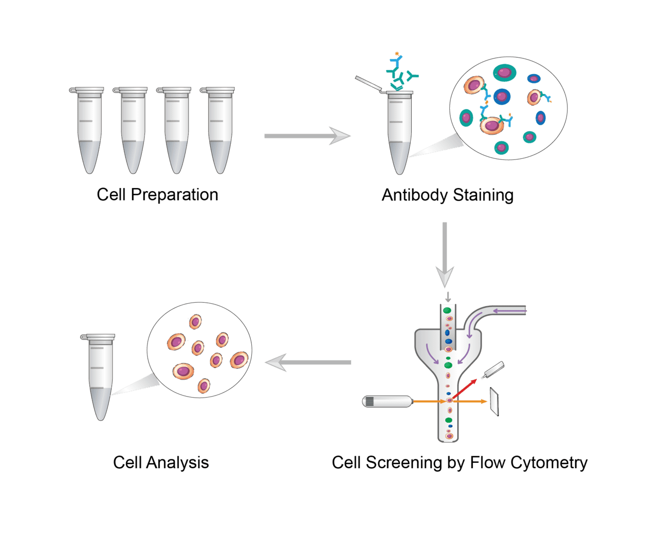
1. Cell Preparation
Choose the appropriate cell sample preparation protocol based on your starting sample. The final sample is a homogeneous single cell suspension. It should contain no clumps or dead cell debris and have a density of 106-107 cells per ml.
2. Antibody Staining
Perform dye labeling on prepared cell suspensions. Select antibodies against specific cell surface molecules are used. Then incubate the cells with fluorescent dye-coupled antibodies. Common staining protocols include intracellular antigen staining, surface antigen staining, etc. Specific protocols can be developed based on experimental needs. After incubation, wash the cells with buffer.
3. Cell Screening by Flow Cytometry
Resuspend the washed cells in the buffer. The labeled cells are then passed through a flow cytometer. The flow cytometer uses a laser to excite the fluorescent dye and a detector to measure the fluorescence intensity of the dye. Based on the fluorescence intensity, the flow cytometer classifies the cells into different groups.
4. Cell Analysis
After screening, target cell populations are collected in test tubes or plates. Next you can check cell purity or other cell analysis using microscopy or flow cytometry.
Troubleshooting
Escapee phenomenon
- Escapers are aggregates of cells (including target cells) that are excluded from flow cytometry analysis, thus altering the results.
- Cell suspension causes. Cell suspensions should be vortexed at low speed. Crude handling such as excessive washing or high speed vortexing can lead to increased cell aggregation.
Collected cells need to be kept alive
- Staining buffer causes. If you need the screened cells to remain viable in subsequent cultures, we recommend that you add serum to the buffer. And avoid containing toxic substances, as this can be toxic to the cells and affect cell viability.
- Staining protocol causes. It is important to note that you may not be able to perform intracellular staining. This is because permeabilization requires disruption of the cell membrane, which can impair cell viability.
- Contamination causes. Experiments should be performed under sterile conditions to ensure that cells are not contaminated.
Are you interested in flow cytometry screening cell protocols? Please contact us for all key points of the relevant protocols, including experimental design, cell sample preparation, antibody selection, and data analysis.
For research use only. Not intended for any clinical use.
Send Inquiry
This site is protected by reCAPTCHA and the Google Privacy Policy and Terms of Service apply.