Live Cell Imaging Protocol & Troubleshooting
Live cell imaging techniques provide clues to the structure and function of cells and tissues. Successful live cell imaging experiments can keep cells in a healthy state on the microscope stage for long periods of time. It also can image a variety of dynamic events with high spatial and temporal resolution over a wide range of time using a variety of instruments.
Creative Biolabs describes the general protocols for live cell imaging, including reagents, procedures and troubleshooting tips for common problems. We help you apply it to examine the structural components of cells, study dynamic processes, molecular localization, and more.
Solutions and Reagents
| Stages | Solutions and Reagents |
| Cell Culture | Culture medium, buffers |
| Cell Labeling | Dyes, fluorescent labeling reagents, background inhibitors, dye stabilizers |
Live cell imaging Procedure
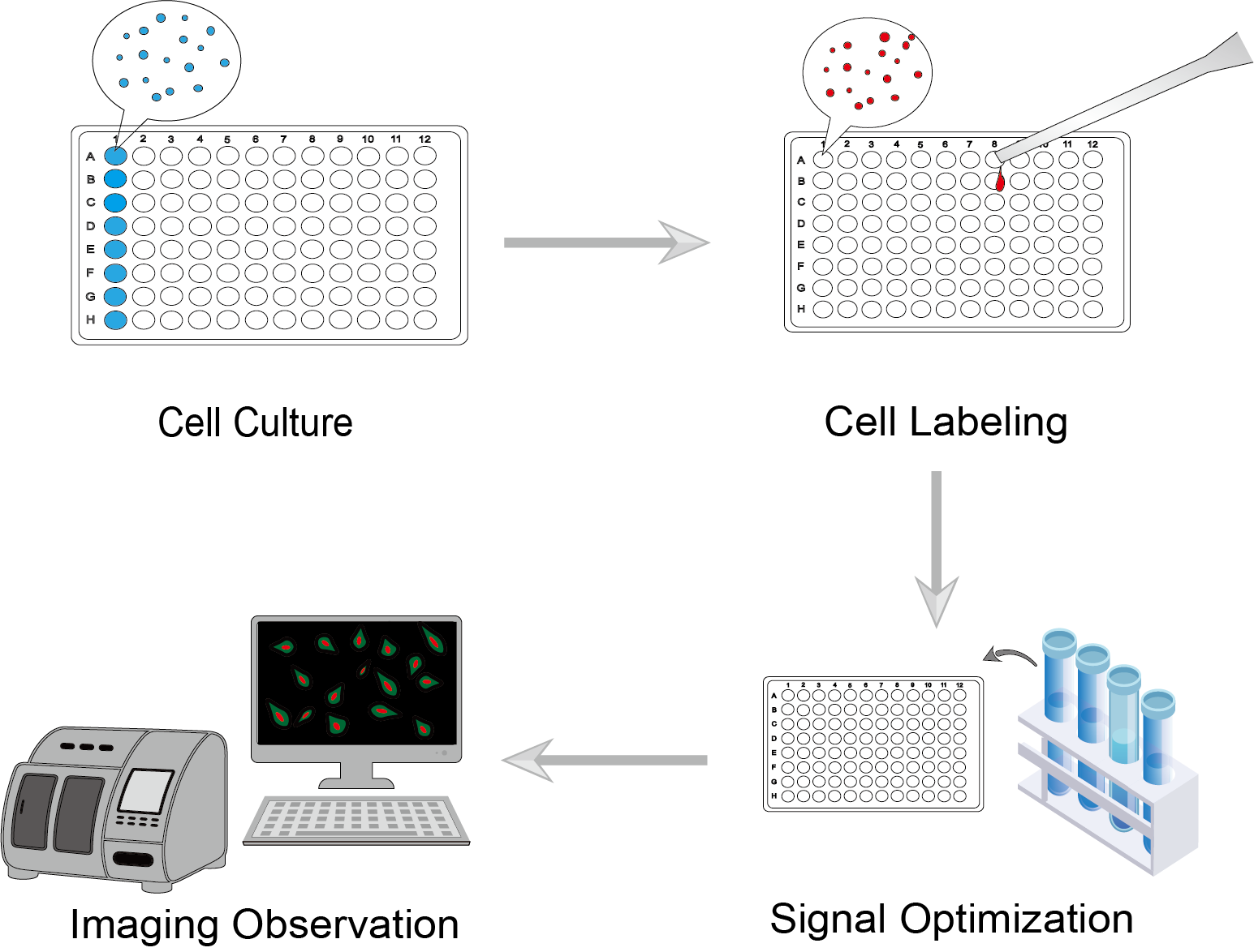
 Figure 1. Live Cell Imaging Procedure.
We need to maintain or culture the cells under optimal conditions. It is not easy to keep cells alive and healthy throughout the imaging observation process. Therefore, the choice of culture medium is particularly important. Cells should be cultured under specific conditions considering physiological temperature, pH, oxygen level, and others.
Select appropriate specific dyes or fluorescent labeling reagents to target cell structure, cell function and target proteins. Avoid using too many markers for the assay.
We recommend using background inhibitors, dye stabilizers and other reagents. Reduce extracellular background fluorescence while being able to maintain cell health, as well as stabilizing markers to reduce signal loss.
Live cell imaging of dynamic processes requires long-term observation. Choose the lowest possible light intensity, exposure time, wavelength range, and excitation energy to illuminate the excited cells, while maintaining a good signal with low background.
Troubleshooting
We list below some common failures and suggested countermeasures to help you improve the success of your live cell imaging.
Low signal
- Marker causes. The possible reason is the unsuitable fluorescent dyes. You can re-select a brighter fluorophore.
- Sample installation causes. The sample is mounted in the incorrect position on the imaging instrument. We recommend using clean coverslips that are sterilized prior to use. Mount the sample as close as possible to the distal end of the coverslip.
- Camera causes. Check the optical instrument settings used. Please use a high numerical aperture, minimally corrected oil-immersion clean objective lens. As well, select a fluorescent filter set that matches the fluorophore. Camera merging can also be used to increase the signal intensity.
High background
- Medium causes. Note that use a phenol red-free medium.
- Labeling causes. To reduce background and improve signal-to-noise ratio, you can use sufficient containment and controls for good antibody staining.
- Other causes. For high background caused by external light, you can turn off the room light and let the light enter through the microscope eyepiece. Choose to use a camera with low readout noise.
Cell damage or death
- Phototoxicity causes. During fluorescence observation, the shorter the wavelength of the excitation light and the more intense the light, the more damage is done to the cells. It will not only weaken the cells but even kill them. We recommend that you use a camera with the highest possible sensitivity. When not capturing images, the excitation light can be turned off. This allows the cells to be exposed to as little excitation light as possible.
- Cell culture environment causes. If the cell environment does not maintain stable and appropriate conditions, the cells may become weakened. You need to use an incubator that consistently provides the right environment, including temperature and humidity. And as much as possible, simulate the environment of standard physiological conditions.
- Contamination causes. Some cells may be affected by bacteria and contaminants that have attached to the container. When performing experiments, you will need to sterilize to ensure that the containers or equipment used are free of bacteria.
Cells out of view
- Cell movement causes. This cell movement cannot be avoided as long as the cells are alive. We recommend that you capture images from multiple locations at the same time, or follow the moving cells while capturing images.
To learn more about the various products and services available for live cell imaging, please visit our related pages.
For research use only. Not intended for any clinical use.
This site is protected by reCAPTCHA and the Google Privacy Policy and Terms of Service apply.

