

Vadastuximab Talirine Overview
Introduction of Vadastuximab Talirine
Vadastuximab talirine (SGN-CD33A) is an antibody-drug conjugate (ADC) directed to CD33 (SIGLEC-3), which is a transmembrane receptor expressed on cells of myeloid lineage. The trial drug is designed for the treatment of acute myeloid leukemia (AML). SGN-CD33A uses h2H12ec as the CD33 antibody. h2H12ec is a humanized IgG1 antibody derived from a murine clone (m2H12; initially raised against the extracellular portion of CD33) that is engineered to contain a cysteine at position 239 of the heavy chain (Ser239Cys) for site-directed drug conjugation. A highly potent, synthetic DNA cross-linking pyrrolobenzodiazepine (PBD) dimer (SGD-1882) is then conjugated to h2H12ec via a protease-cleavable maleimidocaproyl-valine-alanine linker. PBDs are a class of natural products produced by various actinomycetes, are sequence selective DNA alkylating compounds with significant antitumor properties. As a class of DNA-crosslinking agents, they are significantly more potent than systemic chemotherapeutic drugs. Some PBDs have the ability to recognize and bond to specific sequences of DNA. With antibody engineering, very homogenous drug loading is accomplished, averaging 1.9 PBD moieties per antibody. In preclinical studies, SGN-CD33A was more potent than gemtuzumab ozogamicin (GO) against a panel of human AML cell lines and a series of primary AML samples in vitro as well as in immunodeficient mice in vivo. Unlike GO, SGN-CD33A maintained anti-leukemia activity in the presence of drug transporters in preclinical AML models. Although the drug’s anti-leukemia efficacy is now well established, concerns over toxicity to the liver and normal hematopoietic cells have derailed the clinical development of SGN-CD33A, and its future is uncertain.
Mechanism of Action of Vadastuximab Talirine
The CD33 antigen is a sialic acid-dependent adhesion protein that is specific for myeloid cells. CD33 is expressed in approximately 90% of AML cases, as defined by the presence of the antigen on > 20% of the leukemic blasts but not on normal CD34+ pluripotent hematopoietic stem cells or non-hematopoietic tissues. Vadastuximab talirine targets CD33-expressing cells. Binding to the antigen is followed by endocytosis. The covalent link is cleaved inside lysosomes, allowing PBD release. PBD dimers bind in the minor groove of DNA, where they form covalent aminol crosslinks between the N2 of guanine and the C11 position of the PBD. The resulting PBD-DNA adducts cause replication forks to stall and tumor cells to arrest at the G2-M boundary, ultimately resulting in apoptosis at low nanomolar to picomolar concentrations.
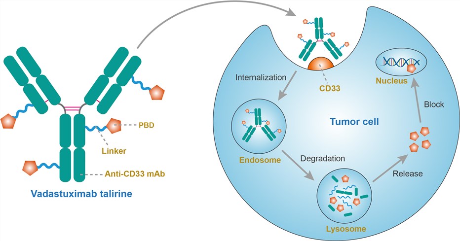 Fig.1 Mechanism of action of vadastuximab talirine
For research use only. Not intended for any clinical use.
Send Inquiry
This site is protected by reCAPTCHA and the Google Privacy Policy and Terms of Service apply.